
Search Count: 91
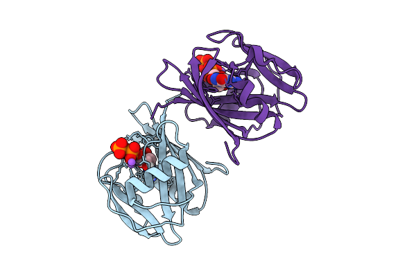 |
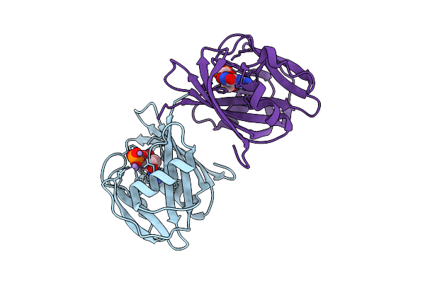
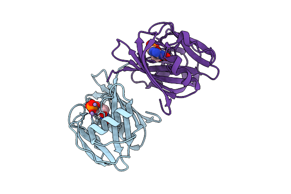
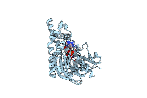
Neutron Crystal Structure Of Human Mth1(G2K/C87A/C104S Mutant) In Complex With 8-Oxo-Dgtp
Organism: Homo sapiens
Method: X-RAY DIFFRACTION, NEUTRON DIFFRACTION Release Date: 2025-07-30 Classification: HYDROLASE Ligands: 8DG, NA |
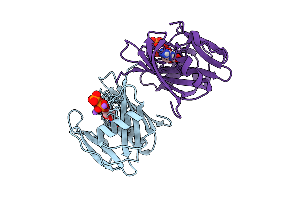 |
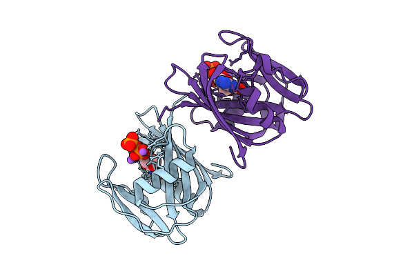
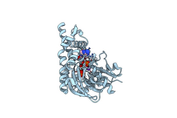
Neutron Crystal Structure Of Human Mth1(G2K/C87A/C104S Mutant) In Complex With 2-Oxo-Datp
Organism: Homo sapiens
Method: X-RAY DIFFRACTION, NEUTRON DIFFRACTION Release Date: 2025-07-30 Classification: HYDROLASE Ligands: 6U4, NA |
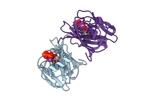 |
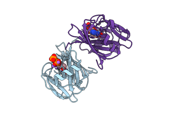
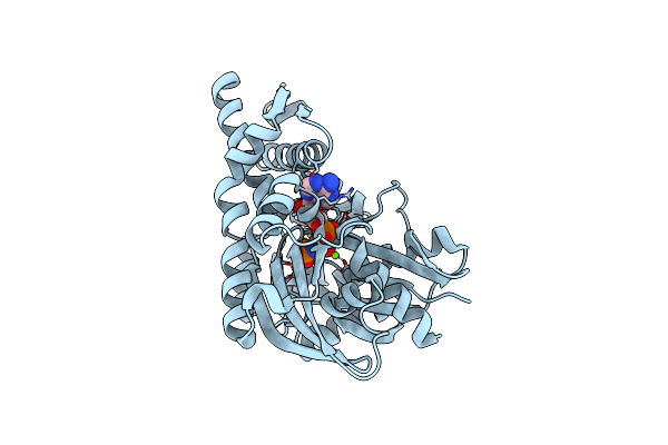
8-Oxo-Dgtp Hydrolysis In Human Mth1(G2K Mutant) Crystal Using Mn2+: The Es Complex
Organism: Homo sapiens
Method: X-RAY DIFFRACTION Release Date: 2025-07-30 Classification: HYDROLASE Ligands: 8DG, NA |
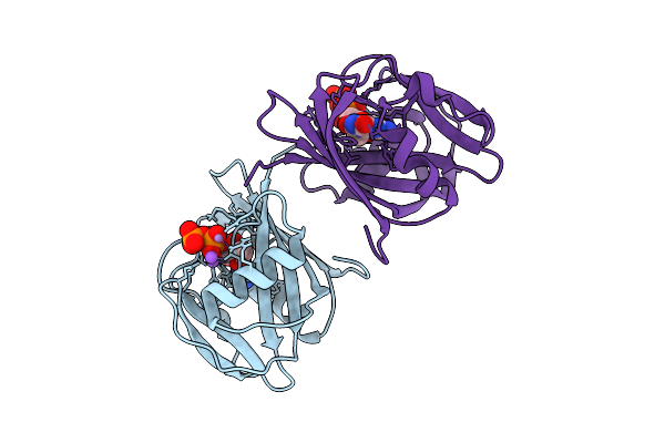 |
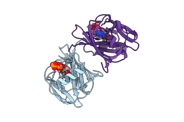
8-Oxo-Dgtp Hydrolysis In Human Mth1(G2K Mutant) Crystal Using Mn2+: The Es-3M Complex
Organism: Homo sapiens
Method: X-RAY DIFFRACTION Release Date: 2025-07-30 Classification: HYDROLASE Ligands: 8DG, NA, MN |
 |
8-Oxo-Dgtp Hydrolysis In Human Mth1(G2K Mutant) Crystal Using Mn2+: The Es/Ep-3M Complex
Organism: Homo sapiens
Method: X-RAY DIFFRACTION Release Date: 2025-07-30 Classification: HYDROLASE Ligands: MN, 8DG, 8OG |
 |
8-Oxo-Dgtp Hydrolysis In Human Mth1(G2K Mutant) Crystal Using Mn2+: The Ep-M Complex
Organism: Homo sapiens
Method: X-RAY DIFFRACTION Release Date: 2025-07-30 Classification: HYDROLASE Ligands: 8OG, MN |
 |
2-Oxo-Datp Hydrolysis In Human Mth1(G2K Mutant) Crystal Using Mn2+: The Es Complex
Organism: Homo sapiens
Method: X-RAY DIFFRACTION Release Date: 2025-07-30 Classification: HYDROLASE Ligands: 6U4, NA |
 |
2-Oxo-Datp Hydrolysis In Human Mth1(G2K Mutant) Crystal Using Mn2+: The Es-2M Complex
Organism: Homo sapiens
Method: X-RAY DIFFRACTION Release Date: 2025-07-30 Classification: HYDROLASE Ligands: 6U4, MN, NA |
 |
2-Oxo-Datp Hydrolysis In Human Mth1(G2K Mutant) Crystal Using Mn2+: The Es/Ep-3M Complex
Organism: Homo sapiens
Method: X-RAY DIFFRACTION Release Date: 2025-07-30 Classification: HYDROLASE Ligands: 6U4, IGU, MN |
 |
2-Oxo-Datp Hydrolysis In Human Mth1(G2K Mutant) Crystal Using Mn2+: The Ep-M Complex
Organism: Homo sapiens
Method: X-RAY DIFFRACTION Release Date: 2025-07-30 Classification: HYDROLASE Ligands: IGU, MN |
 |
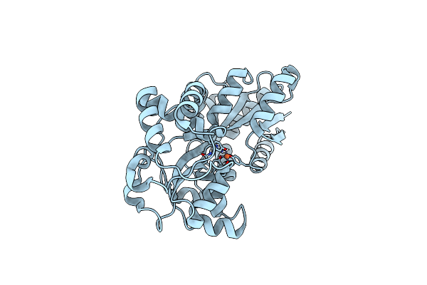
Structure Of Human Delta-1-Pyrroline-5-Carboxylate Dehydrogenase (Aldh4A1) Complexed With The Molecular Tweezer Clr01
Organism: Homo sapiens
Method: X-RAY DIFFRACTION Resolution:2.60 Å Release Date: 2024-05-08 Classification: OXIDOREDUCTASE Ligands: 9SZ |
 |
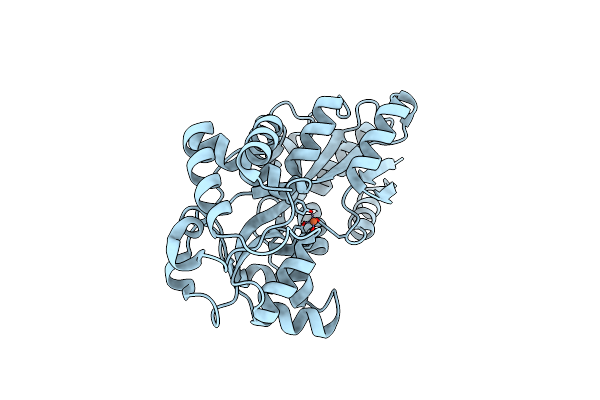
Structure Of Human Delta-1-Pyrroline-5-Carboxylate Dehydrogenase (Aldh4A1) Complexed With A Monophosphate-Tweezer
Organism: Homo sapiens
Method: X-RAY DIFFRACTION Resolution:1.20 Å Release Date: 2024-05-08 Classification: OXIDOREDUCTASE Ligands: A1H1O, BEZ |
 |
Organism: Prochlorococcus marinus subsp. pastoris str. ccmp1986
Method: NEUTRON DIFFRACTION Resolution:2.10 Å Release Date: 2024-01-17 Classification: METAL BINDING PROTEIN Ligands: FE |
 |
Organism: Prochlorococcus marinus subsp. pastoris str. ccmp1986
Method: X-RAY DIFFRACTION Resolution:1.60 Å Release Date: 2023-08-30 Classification: METAL BINDING PROTEIN Ligands: FE |
 |
Organism: Prochlorococcus marinus subsp. pastoris str. ccmp1986
Method: X-RAY DIFFRACTION Resolution:1.65 Å Release Date: 2023-08-30 Classification: METAL BINDING PROTEIN Ligands: FE |
 |
Organism: Prochlorococcus marinus subsp. pastoris str. ccmp1986
Method: X-RAY DIFFRACTION Resolution:1.70 Å Release Date: 2023-08-30 Classification: METAL BINDING PROTEIN Ligands: FE2 |
 |
Organism: Prochlorococcus marinus subsp. pastoris str. ccmp1986
Method: X-RAY DIFFRACTION Resolution:1.76 Å Release Date: 2023-08-30 Classification: METAL BINDING PROTEIN Ligands: FE |
 |
Joint Neutron And X-Ray Crystal Structure Of The Nucleotide-Binding Domain Of Hsp72 In Complex With Adp
Organism: Homo sapiens
Method: X-RAY DIFFRACTION, NEUTRON DIFFRACTION Resolution:1.600 Å, 2.199 Å Release Date: 2022-06-29 Classification: HYDROLASE Ligands: ADP, MG, NA |
 |
Organism: Homo sapiens
Method: X-RAY DIFFRACTION Resolution:1.80 Å Release Date: 2022-06-29 Classification: HYDROLASE Ligands: ADP, MG, CL, K, PO4 |
 |
Organism: Homo sapiens
Method: X-RAY DIFFRACTION Resolution:1.70 Å Release Date: 2022-06-29 Classification: HYDROLASE Ligands: MG, CL, ANP |