
Search Count: 14
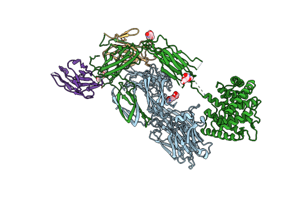 |
Organism: Lama glama, Homo sapiens
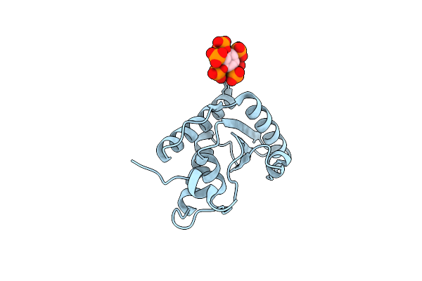
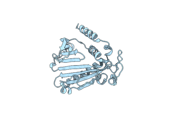
Method: ELECTRON MICROSCOPY Release Date: 2022-03-02 Classification: IMMUNE SYSTEM Ligands: NAG |
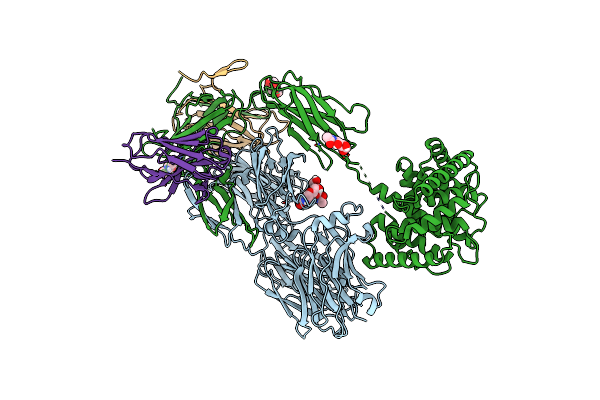 |
Organism: Lama glama, Homo sapiens
Method: ELECTRON MICROSCOPY Release Date: 2022-03-02 Classification: IMMUNE SYSTEM Ligands: NAG |
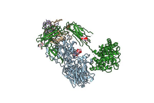 |
Organism: Lama glama, Homo sapiens
Method: ELECTRON MICROSCOPY Release Date: 2022-03-02 Classification: IMMUNE SYSTEM Ligands: NAG |
 |
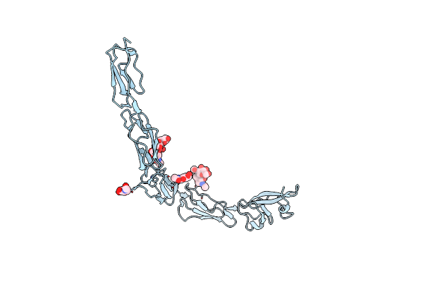
Crystal Structure Of Porcine Hemagglutinating Encephalomyelitis Virus Spike Protein Lectin Domain
Organism: Porcine hemagglutinating encephalomyelitis virus
Method: X-RAY DIFFRACTION Resolution:2.97 Å Release Date: 2019-02-06 Classification: VIRAL PROTEIN Ligands: NAG |
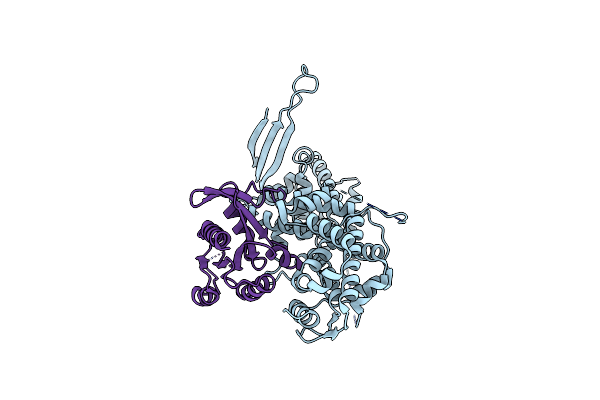 |
Structural Basis For The Selectivity Of Guanine Nucleotide Exchange Factors For The Small G-Protein Ral
Organism: Mus musculus, Drosophila melanogaster
Method: X-RAY DIFFRACTION Resolution:2.60 Å Release Date: 2016-01-13 Classification: SIGNALING PROTEIN |
 |
Structural Basis For The Selectivity Of Guanine Nucleotide Exchange Factors For The Small G-Protein Ral
Organism: Mus musculus, Drosophila melanogaster
Method: X-RAY DIFFRACTION Resolution:2.60 Å Release Date: 2016-01-13 Classification: SIGNALING PROTEIN |
 |
Organism: Homo sapiens
Method: X-RAY DIFFRACTION Resolution:1.50 Å Release Date: 2012-08-08 Classification: LIPID BINDING PROTEIN Ligands: IHP |
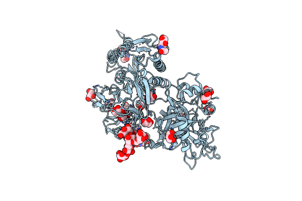 |
Organism: Homo sapiens
Method: X-RAY DIFFRACTION Resolution:2.30 Å Release Date: 2007-02-27 Classification: HYDROLASE Ligands: NAG, GOL |
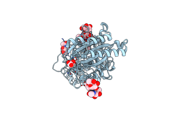 |
Organism: Homo sapiens
Method: X-RAY DIFFRACTION Resolution:2.10 Å Release Date: 2006-10-17 Classification: HYDROLASE Ligands: NAG, MN, MLI |
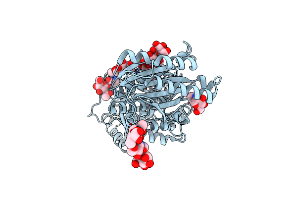 |
Organism: Homo sapiens
Method: X-RAY DIFFRACTION Resolution:2.70 Å Release Date: 2006-10-17 Classification: HYDROLASE Ligands: NAG |
 |
Crystal Structure Of The Drosophila Glutathione S-Transferase-2 In Complex With Glutathione
Organism: Drosophila melanogaster
Method: X-RAY DIFFRACTION Resolution:1.75 Å Release Date: 2003-02-11 Classification: TRANSFERASE Ligands: SO4, GSH |
 |
Crystal Structure Of Mouse Pitp Alpha Void Of Bound Phospholipid At 2.0 Angstroms Resolution
Organism: Mus musculus
Method: X-RAY DIFFRACTION Resolution:2.00 Å Release Date: 2002-05-08 Classification: LIPID BINDING PROTEIN |
 |
Organism: Haementeria officinalis
Method: X-RAY DIFFRACTION Resolution:2.20 Å Release Date: 2001-08-08 Classification: TOXIN Ligands: ROP |
 |
Crystal Structure Of The Glycosylated Five-Domain Human Beta2-Glycoprotein I Purified From Blood Plasma
Organism: Homo sapiens
Method: X-RAY DIFFRACTION Resolution:2.70 Å Release Date: 1999-10-08 Classification: MEMBRANE ADHESION Ligands: NAG |