
Search Count: 27
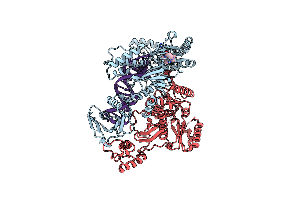 |
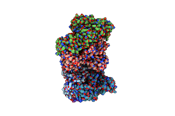
Cryo-Em Structure Of Hiv-1 Reverse Transcriptase With A Dna Aptamer In Complex With Nevirapine
Organism: Human immunodeficiency virus type 1 bh10, Synthetic construct
Method: ELECTRON MICROSCOPY Release Date: 2022-07-20 Classification: TRANSFERASE Ligands: NVP |
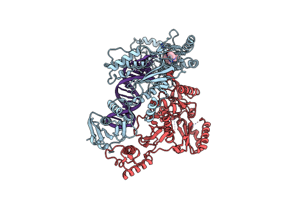 |
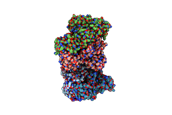
Cryo-Em Structure Of Nnrti Resistant M184I/E138K Mutant Hiv-1 Reverse Transcriptase With A Dna Aptamer In Complex With Nevirapine
Organism: Human immunodeficiency virus type 1 bh10, Synthetic construct
Method: ELECTRON MICROSCOPY Release Date: 2022-07-20 Classification: TRANSFERASE Ligands: NVP |
 |
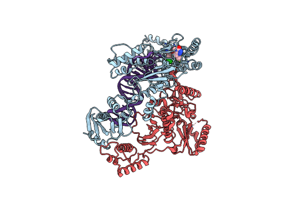
Cryo-Em Structure Of Hiv-1 Reverse Transcriptase With A Dna Aptamer In Complex With Rilpivirine
Organism: Human immunodeficiency virus type 1 bh10, Synthetic construct
Method: ELECTRON MICROSCOPY Release Date: 2022-07-20 Classification: TRANSFERASE Ligands: T27 |
 |
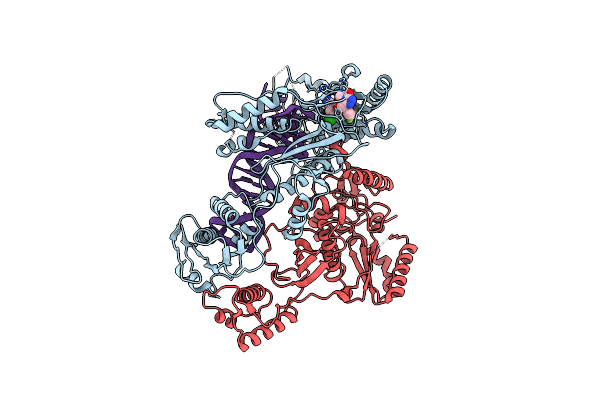
Cryo-Em Structure Of Nnrti Resistant M184I/E138K Mutant Hiv-1 Reverse Transcriptase With A Dna Aptamer In Complex With Rilpivirine
Organism: Human immunodeficiency virus type 1 bh10, Synthetic construct
Method: ELECTRON MICROSCOPY Release Date: 2022-07-20 Classification: TRANSFERASE Ligands: T27 |
 |
Cryo-Em Structure Of Hiv-1 Reverse Transcriptase With A Dna Aptamer In Complex With Doravirine
Organism: Human immunodeficiency virus type 1 bh10, Synthetic construct
Method: ELECTRON MICROSCOPY Release Date: 2022-07-20 Classification: TRANSFERASE Ligands: 2KW |
 |
Cryo-Em Structure Of Nnrti Resistant M184I/E138K Mutant Hiv-1 Reverse Transcriptase With A Dna Aptamer In Complex With Doravirine
Organism: Human immunodeficiency virus type 1 bh10, Synthetic construct
Method: ELECTRON MICROSCOPY Release Date: 2022-07-20 Classification: TRANSFERASE Ligands: 2KW |
 |
Organism: Human immunodeficiency virus type 1 group m subtype b (isolate bh10), Human immunodeficiency virus type 1 bh10, Homo sapiens
Method: X-RAY DIFFRACTION Resolution:3.20 Å Release Date: 2021-12-08 Classification: TRANSFERASE Ligands: MG, 1I4 |
 |
Organism: Human immunodeficiency virus type 1 group m subtype b (isolate bh10), Human immunodeficiency virus type 1 bh10, Homo sapiens
Method: X-RAY DIFFRACTION Resolution:3.00 Å Release Date: 2021-12-08 Classification: TRANSFERASE Ligands: MG, 1I1 |
 |
Organism: Human immunodeficiency virus type 1 group m subtype b (isolate bh10), Human immunodeficiency virus type 1 bh10, Homo sapiens
Method: X-RAY DIFFRACTION Resolution:2.95 Å Release Date: 2021-12-08 Classification: TRANSFERASE Ligands: MG, 1IA |
 |
Organism: Human immunodeficiency virus type 1 group m subtype b (isolate bh10), Human immunodeficiency virus type 1 bh10, Homo sapiens
Method: X-RAY DIFFRACTION Resolution:3.40 Å Release Date: 2021-12-08 Classification: TRANSFERASE Ligands: MN, 1IH |
 |
Organism: Human immunodeficiency virus type 1 group m subtype b (isolate bh10), Human immunodeficiency virus type 1 bh10, Homo sapiens
Method: X-RAY DIFFRACTION Resolution:3.45 Å Release Date: 2021-12-08 Classification: TRANSFERASE Ligands: MG, 1IK |
 |
Organism: Human immunodeficiency virus type 1 group m subtype b (isolate bh10), Human immunodeficiency virus type 1 bh10, Homo sapiens
Method: X-RAY DIFFRACTION Resolution:3.10 Å Release Date: 2021-12-08 Classification: TRANSFERASE Ligands: MG, 1IO |
 |
Organism: Human immunodeficiency virus type 1 group m subtype b (isolate bh10), Human immunodeficiency virus type 1 bh10, Homo sapiens
Method: X-RAY DIFFRACTION Resolution:3.20 Å Release Date: 2021-12-08 Classification: TRANSFERASE Ligands: MG, 1KK |
 |
Crystal Structure Of Hiv-1 Reverse Transcriptase With A Double Stranded Dna In Complex With Fragment 048 At The Transient P-Pocket.
Organism: Human immunodeficiency virus type 1 group m subtype b (isolate bh10), Human immunodeficiency virus type 1 bh10, Homo sapiens
Method: X-RAY DIFFRACTION Resolution:3.30 Å Release Date: 2021-12-08 Classification: TRANSFERASE Ligands: CD, 2NU |
 |
Crystal Structure Of Hiv-1 Reverse Transcriptase With A Double Stranded Dna Showing A Transient P-Pocket
Organism: Human immunodeficiency virus type 1 group m subtype b (isolate bh10), Human immunodeficiency virus type 1 bh10, Homo sapiens
Method: X-RAY DIFFRACTION Resolution:2.85 Å Release Date: 2021-12-08 Classification: TRANSFERASE Ligands: CD, SO4, MG |
 |
Crystal Structure Of Hiv-1 Reverse Transcriptase With A Double Stranded Dna In Complex With Fragment 166 At The Transient P-Pocket.
Organism: Human immunodeficiency virus type 1 group m subtype b (isolate bh10), Human immunodeficiency virus type 1 bh10, Homo sapiens
Method: X-RAY DIFFRACTION Resolution:3.37 Å Release Date: 2021-12-08 Classification: TRANSFERASE Ligands: CD, 3IR |
 |
Cryo-Em Structure Of Hiv-1 Reverse Transcriptase With A Dna Aptamer In Complex With Fragment 166 At The Transient P-Pocket
Organism: Human immunodeficiency virus type 1 group m subtype b (isolate bh10), Human immunodeficiency virus type 1 bh10, Synthetic construct
Method: ELECTRON MICROSCOPY Release Date: 2021-12-08 Classification: TRANSFERASE Ligands: 3IR |
 |
Cryo-Em Structure Of Hiv-1 Reverse Transcriptase With A Dna Aptamer In Complex With Fragment F04 At The Transient P-Pocket
Organism: Human immunodeficiency virus type 1 bh10, Synthetic construct
Method: ELECTRON MICROSCOPY Release Date: 2021-12-08 Classification: TRANSFERASE Ligands: 4OI |
 |
Organism: Musa acuminata
Method: X-RAY DIFFRACTION Resolution:1.51 Å Release Date: 2021-01-27 Classification: SUGAR BINDING PROTEIN Ligands: EDO |
 |
Organism: Musa acuminata
Method: X-RAY DIFFRACTION Resolution:1.80 Å Release Date: 2021-01-27 Classification: SUGAR BINDING PROTEIN Ligands: SO4 |