
Search Count: 23
 |
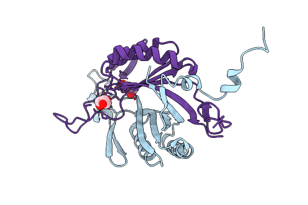
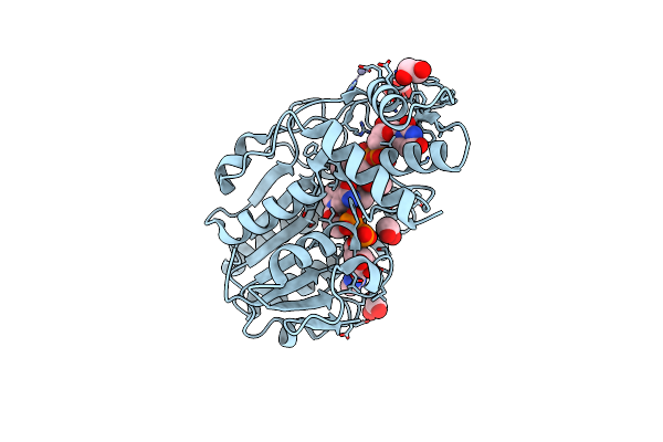
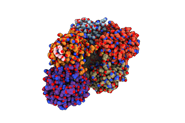
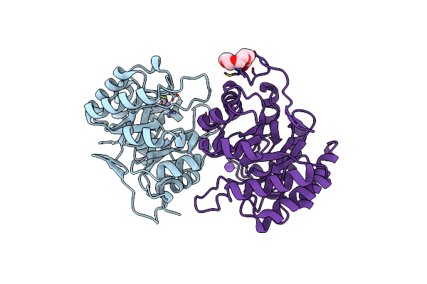
Structure Of Epimerase Mth373 From The Thermophilic Pseudomurein-Containing Methanogen Methanothermobacter Thermautotrophicus
Organism: Methanothermobacter thermautotrophicus str. delta h
Method: X-RAY DIFFRACTION Resolution:2.00 Å Release Date: 2025-02-26 Classification: SUGAR BINDING PROTEIN Ligands: UDP, NAD, EDO, MG, CL |
 |
Organism: Methanothermobacter thermautotrophicus str. delta h
Method: X-RAY DIFFRACTION Resolution:1.97 Å Release Date: 2025-01-29 Classification: SUGAR BINDING PROTEIN Ligands: NAD, EDO, UDX, UDP, GOL |
 |
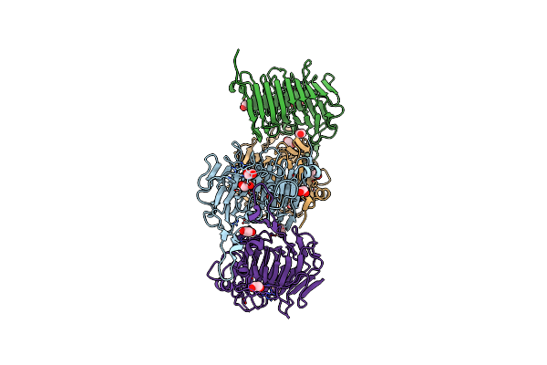
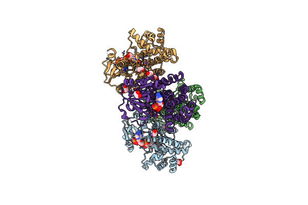
Crystal Structure Of Bacterial Pectin Methylesterase Pmec5 From B. Fibrisolvens D1T
Organism: Butyrivibrio fibrisolvens dsm 3071
Method: X-RAY DIFFRACTION Resolution:1.33 Å Release Date: 2025-01-15 Classification: SUGAR BINDING PROTEIN Ligands: MG, NA, EDO, NH4 |
 |
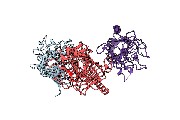
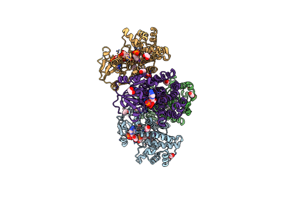
Structure Of Mcrd (Methyl-Coenzyme M Reductase Operon Protein D) From Methanomassiliicoccus Luminyensis
Organism: Methanomassiliicoccus luminyensis b10
Method: X-RAY DIFFRACTION Resolution:1.65 Å Release Date: 2024-07-03 Classification: UNKNOWN FUNCTION Ligands: GOL |
 |
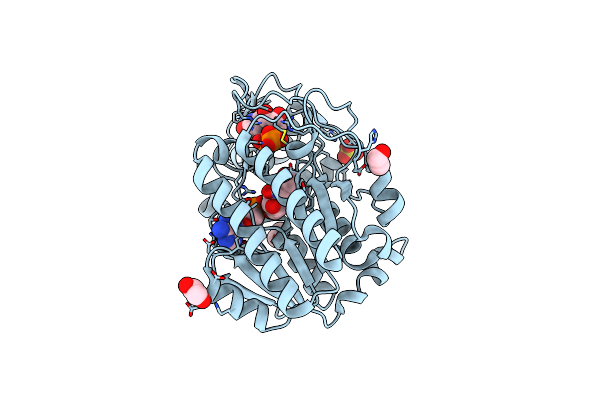
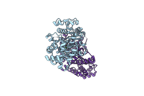
Crystal Structure Of Bacterial Pectin Methylesterase Pmec2 From Rumen Butyrivibrio
Organism: Butyrivibrio fibrisolvens
Method: X-RAY DIFFRACTION Resolution:2.30 Å Release Date: 2023-08-16 Classification: SUGAR BINDING PROTEIN Ligands: CL, EDO |
 |
Crystal Structure Of Bacterial Pectin Methylesterase Pme8A From Rumen Butyrivibrio
Organism: Butyrivibrio proteoclasticus
Method: X-RAY DIFFRACTION Resolution:2.30 Å Release Date: 2023-08-16 Classification: SUGAR BINDING PROTEIN Ligands: EDO |
 |
Structure Of A Pseudomurein Peptide Ligase Type E From Methanothermobacter Thermautotrophicus
Organism: Methanothermobacter thermautotrophicus
Method: X-RAY DIFFRACTION Resolution:2.91 Å Release Date: 2022-10-12 Classification: LIGASE |
 |
Structure Of A Pseudomurein Peptide Ligase Type E From Methanothermus Fervidus
Organism: Methanothermus fervidus
Method: X-RAY DIFFRACTION Resolution:2.00 Å Release Date: 2022-10-12 Classification: LIGASE Ligands: UDP, SO4 |
 |
Apo Structure Of A Pseudomurein Peptide Ligase Type E From Methanothermus Fervidus
Organism: Methanothermus fervidus (strain atcc 43054 / dsm 2088 / jcm 10308 / v24 s)
Method: X-RAY DIFFRACTION Resolution:1.84 Å Release Date: 2021-09-01 Classification: LIGASE Ligands: MG, SO4 |
 |
Structure Of A Pseudomurein Peptide Ligase Type E From Methanothermus Fervidus
Organism: Methanothermus fervidus
Method: X-RAY DIFFRACTION Resolution:1.90 Å Release Date: 2021-08-11 Classification: LIGASE Ligands: PO4, MG, EDO, SO4, UDP |
 |
Structure Of A Pseudomurein Peptide Ligase Type C From Methanothermus Fervidus
Organism: Methanothermus fervidus
Method: X-RAY DIFFRACTION Resolution:2.50 Å Release Date: 2021-02-10 Classification: LIGASE Ligands: SO4, ZN, GOL, AAE |
 |
Structure Of Epimerase Mth375 From The Thermophilic Pseudomurein-Containing Methanogen Methanothermobacter Thermautotrophicus
Organism: Methanothermobacter thermautotrophicus (strain atcc 29096 / dsm 1053 / jcm 10044 / nbrc 100330 / delta h)
Method: X-RAY DIFFRACTION Resolution:2.30 Å Release Date: 2020-07-08 Classification: ISOMERASE Ligands: PO4, GOL, CL, UDP, AMP |
 |
Structure Of Epimerase Mth375 From The Thermophilic Pseudomurein-Containing Methanogen Methanothermobacter Thermautotrophicus
Organism: Methanothermobacter thermautotrophicus
Method: X-RAY DIFFRACTION Resolution:2.01 Å Release Date: 2020-07-08 Classification: ISOMERASE Ligands: NAD, UDP, EDO, SO4 |
 |
2,5-Diamino-6-(Ribosylamino)-4(3H)-Pyrimidinone 5'-Phosphate Reductase (Mthred) From Methanothermobacter Thermautotrophicus
Organism: Methanothermobacter thermautotrophicus (strain atcc 29096 / dsm 1053 / jcm 10044 / nbrc 100330 / delta h)
Method: X-RAY DIFFRACTION Resolution:2.07 Å Release Date: 2020-06-10 Classification: OXIDOREDUCTASE Ligands: CL, GOL, MG, NAP |
 |
Udp-N-Acetylglucosamine 4-Epimerase From Methanobrevibacter Ruminantium M1 In Complex With Udp-N-Acetylmuramic Acid
Organism: Methanobrevibacter ruminantium (strain atcc 35063 / dsm 1093 / jcm 13430 / ocm 146 / m1)
Method: X-RAY DIFFRACTION Resolution:1.66 Å Release Date: 2018-10-03 Classification: SUGAR BINDING PROTEIN Ligands: NAD, EPZ, ZN, EDO |
 |
Organism: Methanocaldococcus jannaschii dsm 2661
Method: X-RAY DIFFRACTION Resolution:2.20 Å Release Date: 2015-07-22 Classification: OXIDOREDUCTASE Ligands: NDP, K, ZN, EDO, NA |
 |
Crystal Structure Of The Methanocaldococcus Jannaschii G1Pdh With Nadph And Dhap
Organism: Methanocaldococcus jannaschii
Method: X-RAY DIFFRACTION Resolution:2.23 Å Release Date: 2015-07-22 Classification: OXIDOREDUCTASE Ligands: NDP, K, ZN, 13P, EDO, 1GP |
 |
Organism: Methanocaldococcus jannaschii
Method: X-RAY DIFFRACTION Resolution:2.45 Å Release Date: 2015-07-22 Classification: OXIDOREDUCTASE Ligands: MG, ZN |
 |
Crystal Structure Of Pyrococcus Furiosus 3-Deoxy-D-Arabino- Heptulosonate 7-Phosphate Synthase
Organism: Pyrococcus furiosus
Method: X-RAY DIFFRACTION Resolution:2.15 Å Release Date: 2013-11-20 Classification: TRANSFERASE Ligands: CO3, PEP, CD, CL, 2PE |
 |
Crystal Structure Of Pyrococcus Furiosus 3-Deoxy-D-Arabino- Heptulosonate 7-Phosphate Synthase I181D Interface Mutant
Organism: Pyrococcus furiosus
Method: X-RAY DIFFRACTION Resolution:1.80 Å Release Date: 2013-11-20 Classification: TRANSFERASE Ligands: MN, CL, K, 1PE |