
Search Count: 40
 |
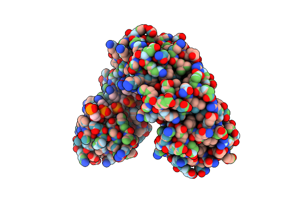
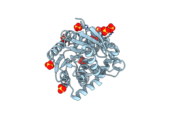
Organism: Canavalia ensiformis
Method: X-RAY DIFFRACTION Resolution:2.69 Å Release Date: 2020-11-11 Classification: PLANT PROTEIN, Hydrolase |
 |
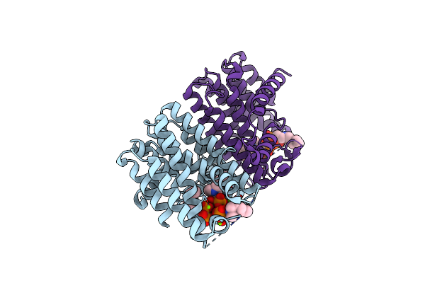
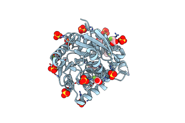
Organism: Canavalia ensiformis
Method: X-RAY DIFFRACTION Resolution:2.10 Å Release Date: 2020-11-11 Classification: PLANT PROTEIN Ligands: MN, CA, ZN, EDO |
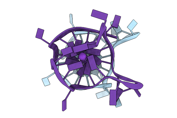 |
Organism: Homo sapiens
Method: X-RAY DIFFRACTION Resolution:1.60 Å Release Date: 2020-04-15 Classification: DNA Ligands: K |
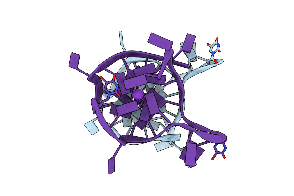 |
Organism: Homo sapiens
Method: X-RAY DIFFRACTION Resolution:1.80 Å Release Date: 2020-04-15 Classification: DNA Ligands: K |
 |
Crystal Structure Of Smt Fusion Peptidyl-Prolyl Cis-Trans Isomerase From Burkholderia Pseudomallei Complexed With Sf339
Organism: Saccharomyces cerevisiae, Burkholderia pseudomallei (strain 1710b)
Method: X-RAY DIFFRACTION Resolution:1.85 Å Release Date: 2020-02-12 Classification: ISOMERASE, PROTEIN BINDING Ligands: LL7, CA, EDO |
 |
Crystal Structure Of Smt Fusion Peptidyl-Prolyl Cis-Trans Isomerase From Burkholderia Pseudomallei Complexed With Sf355
Organism: Saccharomyces cerevisiae, Burkholderia pseudomallei (strain 1710b)
Method: X-RAY DIFFRACTION Resolution:2.10 Å Release Date: 2020-02-12 Classification: ISOMERASE, PROTEIN BINDING Ligands: CA, LLD |
 |
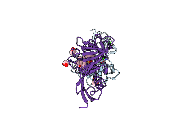
Crystal Structure Of A Designer Pentatrico Peptide Rna Binding Protein, Bound To A Complex Rna Target And Featuring An Infinite Superhelix And Microheterogeneity.
Organism: Zea mays
Method: X-RAY DIFFRACTION Resolution:2.01 Å Release Date: 2019-08-21 Classification: RNA BINDING PROTEIN/RNA |
 |
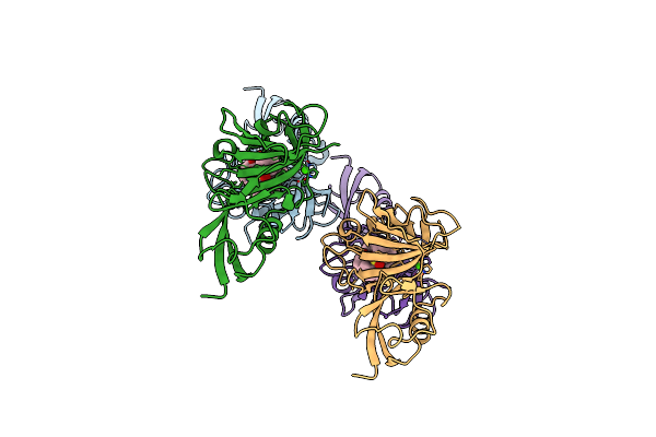
Butelase 1: Auto-Catalytic Cleavage As An Evolutionary Constraint For Macrocyclizing Endopeptidases
Organism: Clitoria ternatea
Method: X-RAY DIFFRACTION Resolution:3.10 Å Release Date: 2018-08-15 Classification: PLANT PROTEIN, Hydrolase |
 |
Asparaginyl Endopeptidase 1 Bound To Aan Peptide, A Tetrahedral Intermediate
Organism: Helianthus annuus, Synthetic construct
Method: X-RAY DIFFRACTION Resolution:1.80 Å Release Date: 2018-02-07 Classification: PLANT PROTEIN Ligands: GOL |
 |
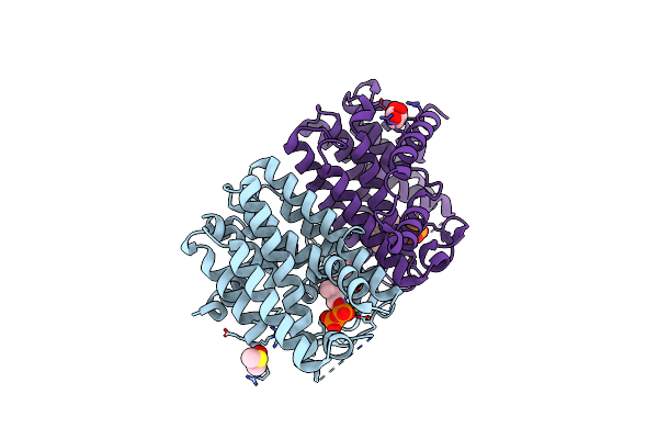
Native Structure Of Farnesyl Pyrophosphate Synthase From Pseudomonas Aeruginosa Pa01, With Bound Ibandronic Acid Molecules.
Organism: Pseudomonas aeruginosa
Method: X-RAY DIFFRACTION Resolution:1.85 Å Release Date: 2015-03-11 Classification: TRANSFERASE Ligands: MG, BFQ |
 |
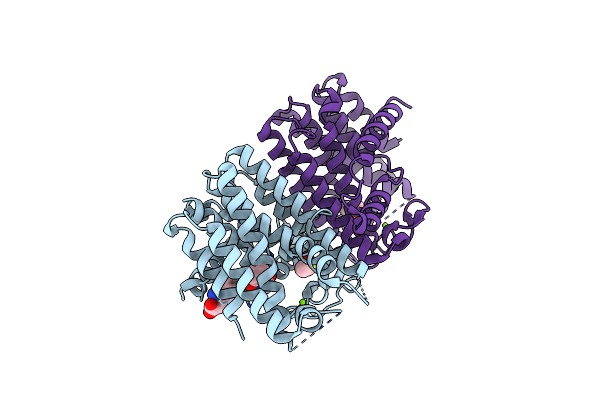
Native Structure Of Farnesyl Pyrophosphate Synthase From Pseudomonas Aeruginosa Pa01, With Bound Fragment Spb02696, And Substrate Geranyl Pyrophosphate.
Organism: Pseudomonas aeruginosa pao1
Method: X-RAY DIFFRACTION Resolution:1.55 Å Release Date: 2014-03-12 Classification: TRANSFERASE Ligands: MG, GPP, 6H6, GOL, DMS |
 |
Native Structure Of Farnesyl Pyrophosphate Synthase From Pseudomonas Aeruginosa Pa01, With Bound Fragment Spb02696.
Organism: Pseudomonas aeruginosa pao1
Method: X-RAY DIFFRACTION Resolution:1.90 Å Release Date: 2014-02-26 Classification: TRANSFERASE Ligands: 6H6, DMS, CL |
 |
Native Structure Of Farnesyl Pyrophosphate Synthase From Pseudomonas Aeruginosa Pa01, With Bound Substrate Molecule Geranyl Pyrophosphate.
Organism: Pseudomonas aeruginosa pao1
Method: X-RAY DIFFRACTION Resolution:1.87 Å Release Date: 2014-02-26 Classification: TRANSFERASE Ligands: GPP, MG, DMS, GOL |
 |
Native Structure Of Farnesyl Pyrophosphate Synthase From Pseudomonas Aeruginosa Pao1, With Bound Fragment Km10833.
Organism: Pseudomonas aeruginosa pao1
Method: X-RAY DIFFRACTION Resolution:1.85 Å Release Date: 2014-02-12 Classification: TRANSFERASE Ligands: DMS, MG, NVU |
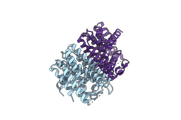 |
Native Structure Of Farnesyl Pyrophosphate Synthase From Pseudomonas Aeruginosa Pa01.
Organism: Pseudomonas aeruginosa pao1
Method: X-RAY DIFFRACTION Resolution:1.55 Å Release Date: 2013-12-04 Classification: TRANSFERASE |
 |
Structure Of A Putative Epoxide Hydrolase Q244E Mutant From Pseudomonas Aeruginosa, With Bound Mfa.
Organism: Pseudomonas aeruginosa pao1
Method: X-RAY DIFFRACTION Resolution:1.77 Å Release Date: 2013-10-09 Classification: HYDROLASE Ligands: SO4, FAH |
 |
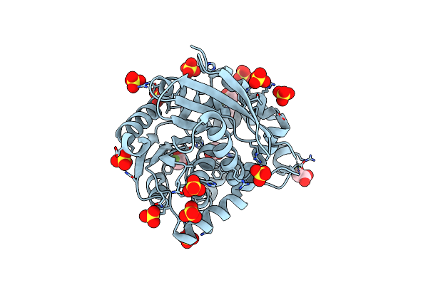
Structure Of A Putative Epoxide Hydrolase T131D Mutant From Pseudomonas Aeruginosa.
Organism: Pseudomonas aeruginosa pao1
Method: X-RAY DIFFRACTION Resolution:1.30 Å Release Date: 2013-10-02 Classification: HYDROLASE Ligands: GOL, SO4, CL |
 |
Structure Of A Putative Epoxide Hydrolase T131D Mutant From Pseudomonas Aeruginosa, With Bound Mfa
Organism: Pseudomonas aeruginosa pao1
Method: X-RAY DIFFRACTION Resolution:1.55 Å Release Date: 2013-10-02 Classification: HYDROLASE Ligands: SO4, FAH, CL |
 |
Structure Of A Putative Epoxide Hydrolase Q244E Mutant From Pseudomonas Aeruginosa.
Organism: Pseudomonas aeruginosa pao1
Method: X-RAY DIFFRACTION Resolution:1.35 Å Release Date: 2013-10-02 Classification: HYDROLASE Ligands: SO4, GOL, CL |
 |
Organism: Pseudomonas aeruginosa pao1
Method: X-RAY DIFFRACTION Release Date: 2013-02-06 Classification: HYDROLASE Ligands: SO4, GOL |