
Search Count: 14
 |
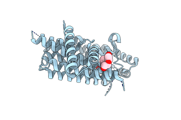
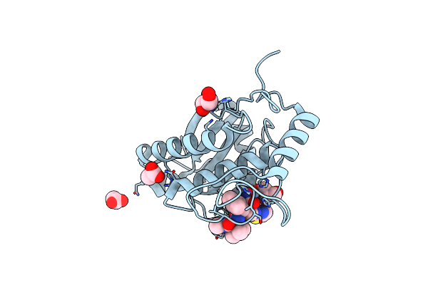
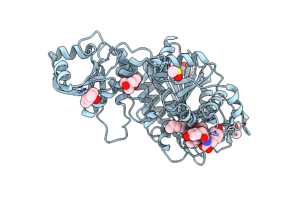
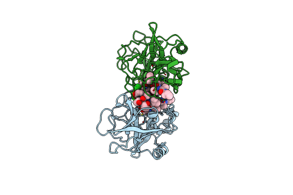
Crystal Structure Of Human Il-1Beta In Complex With A Low Molecular Weight Antagonist
Organism: Homo sapiens
Method: X-RAY DIFFRACTION Resolution:1.95 Å Release Date: 2023-09-13 Classification: CYTOKINE Ligands: T9C |
 |
Organism: Trypanosoma brucei
Method: X-RAY DIFFRACTION Resolution:1.67 Å Release Date: 2020-04-08 Classification: TRANSFERASE Ligands: DMS, PEG |
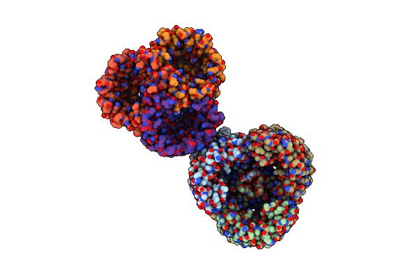 |
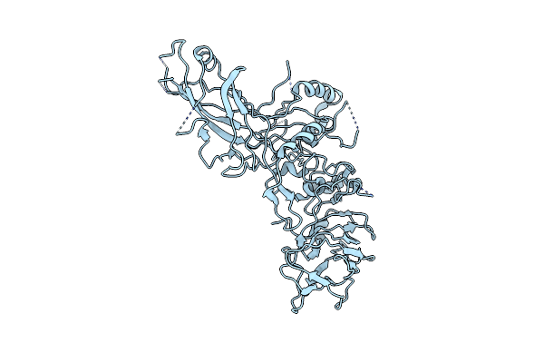
Crystal Structure Of The Outer Membrane Channel Dcap Of Acinetobacter Baumannii
Organism: Acinetobacter baumannii
Method: X-RAY DIFFRACTION Resolution:2.20 Å Release Date: 2018-11-14 Classification: MEMBRANE PROTEIN |
 |
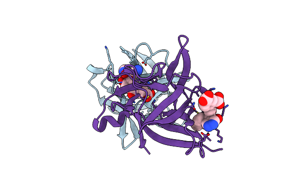
High-Resolution Crystal Structure Of The P-I Snake Venom Metalloproteinase Bap1 In Complex With A Peptidomimetic: Insights Into Inhibitor Binding
Organism: Bothrops asper
Method: X-RAY DIFFRACTION Resolution:1.46 Å Release Date: 2009-06-16 Classification: HYDROLASE/INHIBITOR Ligands: ZN, WR2, GOL |
 |
High-Resolution Crystal Structure Of The P-I Snake Venom Metalloproteinase Bap1 In Complex With A Peptidomimetic: Insights Into Inhibitor Binding
Organism: Bothrops asper
Method: X-RAY DIFFRACTION Resolution:1.14 Å Release Date: 2009-06-16 Classification: HYDROLASE/INHIBITOR Ligands: ZN, WR2, GOL, ACT |
 |
High-Resolution Crystal Structure Of The P-I Snake Venom Metalloproteinase Bap1 In Complex With A Peptidomimetic: Insights Into Inhibitor Binding
Organism: Bothrops asper
Method: X-RAY DIFFRACTION Resolution:1.08 Å Release Date: 2009-06-16 Classification: HYDROLASE/INHIBITOR Ligands: ZN, WR2, GOL, IMD |
 |
High-Resolution Crystal Structure Of The P-I Snake Venom Metalloproteinase Bap1 In Complex With A Peptidomimetic: Insights Into Inhibitor Binding
Organism: Bothrops asper
Method: X-RAY DIFFRACTION Resolution:1.05 Å Release Date: 2009-06-16 Classification: HYDROLASE/INHIBITOR Ligands: ZN, WR2, GOL |
 |
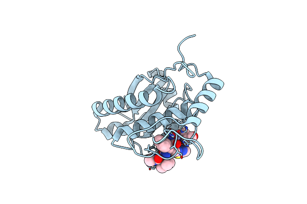
Organism: Arthrobacter nicotinovorans
Method: X-RAY DIFFRACTION Resolution:2.10 Å Release Date: 2007-01-04 Classification: HYDROLASE |
 |
Organism: Bos taurus, Microcystis sp.
Method: X-RAY DIFFRACTION Resolution:1.73 Å Release Date: 2006-12-06 Classification: HYDROLASE/INHIBITOR Ligands: CL, MPD, DMS, ZN, AHY |
 |
Crystal Structure Of The Enzymatic Component C2-I Of The C2-Toxin From Clostridium Botulinum At Ph 3.0
Organism: Clostridium botulinum
Method: X-RAY DIFFRACTION Resolution:2.11 Å Release Date: 2006-10-11 Classification: TOXIN Ligands: GOL, SO4 |
 |
Crystal Structure Of The Enzymatic Component C2-I Of The C2-Toxin From Clostridium Botulinum At Ph 3.0 (Mut-S361R)
Organism: Clostridium botulinum
Method: X-RAY DIFFRACTION Resolution:1.75 Å Release Date: 2006-10-11 Classification: TOXIN Ligands: GOL, SO4 |
 |
Crystal Structure Of The Enzymatic Component C2-I Of The C2-Toxin From Clostridium Botulinum At Ph 6.1
Organism: Clostridium botulinum
Method: X-RAY DIFFRACTION Resolution:2.30 Å Release Date: 2006-10-11 Classification: TOXIN Ligands: CO, GOL, SO4 |
 |
Low Quality Crystal Structure Of The Transport Component C2-Ii Of The C2-Toxin From Clostridium Botulinum
Organism: Clostridium botulinum
Method: X-RAY DIFFRACTION Resolution:3.13 Å Release Date: 2006-10-11 Classification: TOXIN |
 |
Organism: Sus scrofa, Scytonema hofmanni
Method: X-RAY DIFFRACTION Resolution:2.80 Å Release Date: 2003-10-24 Classification: HYDROLASE/HYDROLASE INHIBITOR |