
Search Count: 16
All
Selected
 |
Organism: Homo sapiens
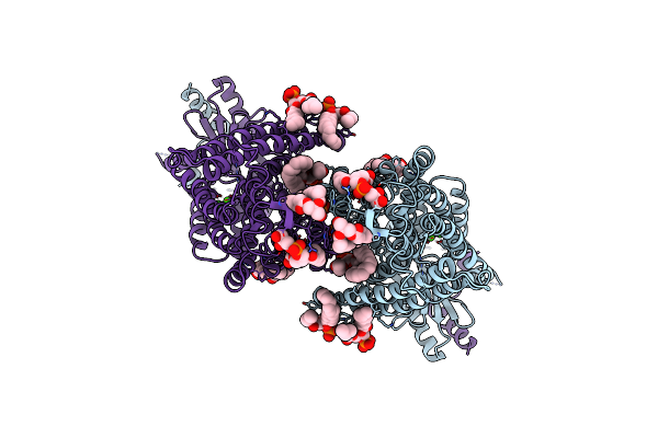
Method: ELECTRON MICROSCOPY Release Date: 2025-05-21 Classification: MEMBRANE PROTEIN |
 |
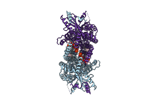
Organism: Homo sapiens
Method: ELECTRON MICROSCOPY Release Date: 2023-09-06 Classification: MEMBRANE PROTEIN |
 |
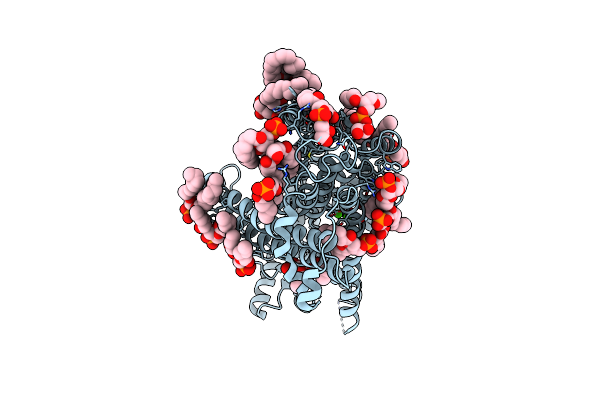
Organism: Homo sapiens
Method: ELECTRON MICROSCOPY Release Date: 2023-09-06 Classification: MEMBRANE PROTEIN |
 |
Aftmem16 In C22 Lipid Nanodiscs With Msp2N2 Scaffold Protein In The Presnece Of Ca2+
Organism: Neosartorya fumigata (strain atcc mya-4609 / af293 / cbs 101355 / fgsc a1100)
Method: ELECTRON MICROSCOPY Release Date: 2022-05-18 Classification: LIPID TRANSPORT Ligands: CA, PGW |
 |
Aftmem16 In C22 Lipid Nanodiscs With Msp1E3 Scaffold Protein In The Presnece Of Ca2+
Organism: Neosartorya fumigata (strain cea10 / cbs 144.89 / fgsc a1163)
Method: ELECTRON MICROSCOPY Release Date: 2022-05-18 Classification: LIPID TRANSPORT Ligands: CA, PGW |
 |
Aftmem16 In C14 Lipid Nanodiscs With Msp1E3 Scaffold Protein In The Absence Of Ca2+
Organism: Neosartorya fumigata (strain cea10 / cbs 144.89 / fgsc a1163)
Method: ELECTRON MICROSCOPY Release Date: 2022-05-18 Classification: LIPID TRANSPORT Ligands: PGW |
 |
Organism: Neosartorya fumigata (strain cea10 / cbs 144.89 / fgsc a1163)
Method: ELECTRON MICROSCOPY Release Date: 2022-05-18 Classification: LIPID TRANSPORT Ligands: PGW |
 |
Organism: Neosartorya fumigata (strain cea10 / cbs 144.89 / fgsc a1163)
Method: ELECTRON MICROSCOPY Release Date: 2022-05-18 Classification: LIPID TRANSPORT Ligands: PGW |
 |
Aftmem16 In C18 Lipid Nanodiscs With Msp1E3 Scaffold Protein In The Presence Of Ca2+, Full Dimer
Organism: Neosartorya fumigata (strain cea10 / cbs 144.89 / fgsc a1163)
Method: ELECTRON MICROSCOPY Release Date: 2022-05-18 Classification: LIPID TRANSPORT Ligands: CA, PGW |
 |
Aftmem16 In C18 Lipid Nanodiscs With Msp1E3 Scaffold Protein In The Presence Of Ca2+, Monomer With Extra Lipids
Organism: Neosartorya fumigata (strain cea10 / cbs 144.89 / fgsc a1163)
Method: ELECTRON MICROSCOPY Release Date: 2022-05-18 Classification: LIPID TRANSPORT Ligands: CA, PGW |
 |
Organism: Mus musculus
Method: X-RAY DIFFRACTION Resolution:2.05 Å Release Date: 2020-02-05 Classification: TOXIN Ligands: GOL, CL |
 |
Organism: Mus musculus
Method: ELECTRON MICROSCOPY Release Date: 2020-02-05 Classification: TOXIN Ligands: NAG |
 |
Organism: Mus musculus
Method: X-RAY DIFFRACTION Resolution:3.17 Å Release Date: 2020-02-05 Classification: TOXIN |
 |
Organism: Mus musculus
Method: ELECTRON MICROSCOPY Release Date: 2020-02-05 Classification: TOXIN Ligands: NAG |
 |
The Electron Crystallography Structure Of The Camp-Bound Potassium Channel Mlok1
Organism: Mesorhizobium loti
Method: ELECTRON CRYSTALLOGRAPHY Resolution:7.00 Å Release Date: 2014-01-15 Classification: TRANSPORT Ligands: K |
 |
The Electron Crystallography Structure Of The Camp-Free Potassium Channel Mlok1
Organism: Mesorhizobium loti
Method: ELECTRON CRYSTALLOGRAPHY Resolution:7.00 Å Release Date: 2014-01-15 Classification: TRANSPORT PROTEIN Ligands: K |