
Search Count: 110
 |
Organism: Homo sapiens
Method: X-RAY DIFFRACTION Release Date: 2025-03-12 Classification: LIGASE Ligands: A1I41, EDO |
 |
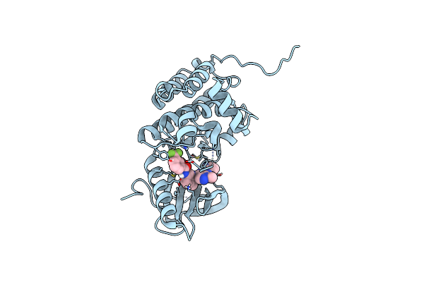
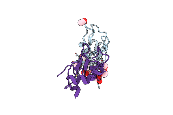
Crystal Structure Of Human Trim7 Pryspry Domain Bound To (2-(1-Oxoisoindolin-2-Yl)-3-Phenylpropanoyl)-L-Glutamine
Organism: Homo sapiens
Method: X-RAY DIFFRACTION Resolution:1.60 Å Release Date: 2024-01-17 Classification: LIGASE Ligands: Y3C, EDO, SRT |
 |
Organism: Homo sapiens
Method: X-RAY DIFFRACTION Resolution:1.80 Å Release Date: 2024-01-17 Classification: LIGASE Ligands: MLA, EDO |
 |
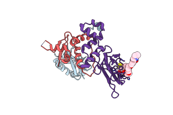
Crystal Structure Of Ephrin A2 (Epha2) Receptor Protein Kinase With Compound 19
Organism: Homo sapiens
Method: X-RAY DIFFRACTION Resolution:1.90 Å Release Date: 2023-11-29 Classification: TRANSFERASE Ligands: QUF |
 |
Crystal Structure Of Human Ephrin Type-A Receptor 2 (Epha2) Kinase Domain In Complex With Jg165
Organism: Homo sapiens
Method: X-RAY DIFFRACTION Resolution:1.80 Å Release Date: 2023-11-22 Classification: TRANSFERASE Ligands: WT3, EDO |
 |
Crystal Structure Of Ephrin A2 (Epha2) Receptor Protein Kinase With Compound 20
Organism: Homo sapiens
Method: X-RAY DIFFRACTION Resolution:1.50 Å Release Date: 2023-03-08 Classification: TRANSFERASE Ligands: QTX |
 |
Crystal Structure Of Ephrin A2 (Epha2) Receptor Protein Kinase With Compound 12
Organism: Homo sapiens
Method: X-RAY DIFFRACTION Resolution:1.82 Å Release Date: 2023-03-08 Classification: TRANSFERASE Ligands: R3L |
 |
Crystal Structure Of Ephrin A2 (Epha2) Receptor Protein Kinase With Compound 7
Organism: Homo sapiens
Method: X-RAY DIFFRACTION Resolution:1.47 Å Release Date: 2023-03-08 Classification: TRANSFERASE Ligands: QUU |
 |
Crystal Structure Of Ephrin A2 (Epha2) Receptor Protein Kinase With Compound 8
Organism: Homo sapiens
Method: X-RAY DIFFRACTION Resolution:1.42 Å Release Date: 2023-03-08 Classification: TRANSFERASE Ligands: R0T |
 |
Crystal Structure Of Ephrin A2 (Epha2) Receptor Protein Kinase With Compound 9
Organism: Homo sapiens
Method: X-RAY DIFFRACTION Resolution:1.63 Å Release Date: 2023-03-08 Classification: TRANSFERASE Ligands: R0O |
 |
Crystal Structure Of Ephrin A2 (Epha2) Receptor Protein Kinase With Compound 11
Organism: Homo sapiens
Method: X-RAY DIFFRACTION Resolution:2.02 Å Release Date: 2023-03-08 Classification: TRANSFERASE Ligands: R0X |
 |
Crystal Structure Of Ephrin A2 (Epha2) Receptor Protein Kinase With Compound 14
Organism: Homo sapiens
Method: X-RAY DIFFRACTION Resolution:1.12 Å Release Date: 2023-03-08 Classification: TRANSFERASE Ligands: QU6 |
 |
Organism: Homo sapiens
Method: X-RAY DIFFRACTION Resolution:2.30 Å Release Date: 2022-12-28 Classification: LIGASE Ligands: SYF |
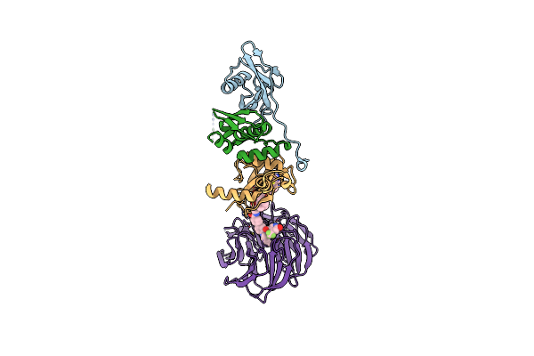 |
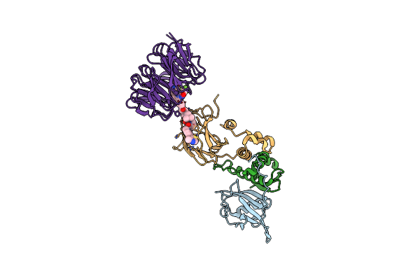
Structure Of Human Wdr5 And Pvhl:Elonginc:Elonginb Bound To Protac With C3 Linker
Organism: Homo sapiens
Method: X-RAY DIFFRACTION Resolution:2.80 Å Release Date: 2022-11-09 Classification: LIGASE Ligands: Q3R |
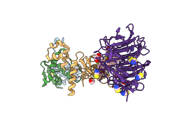 |
Structure Of Human Wdr5 And Pvhl:Elonginc:Elonginb Bound To Protac With Aryl Linker
Organism: Homo sapiens
Method: X-RAY DIFFRACTION Resolution:2.20 Å Release Date: 2022-11-09 Classification: LIGASE Ligands: SCN, Q43, EDO, K |
 |
Organism: Homo sapiens
Method: X-RAY DIFFRACTION Resolution:1.30 Å Release Date: 2022-02-09 Classification: LIGASE Ligands: ZN, EDO |
 |
Quaternary Complex Of Human Wdr5 And Pvhl:Elonginc:Elonginb Bound To Protac Homer
Organism: Homo sapiens
Method: X-RAY DIFFRACTION Resolution:2.50 Å Release Date: 2021-11-24 Classification: STRUCTURAL PROTEIN Ligands: 8KH |
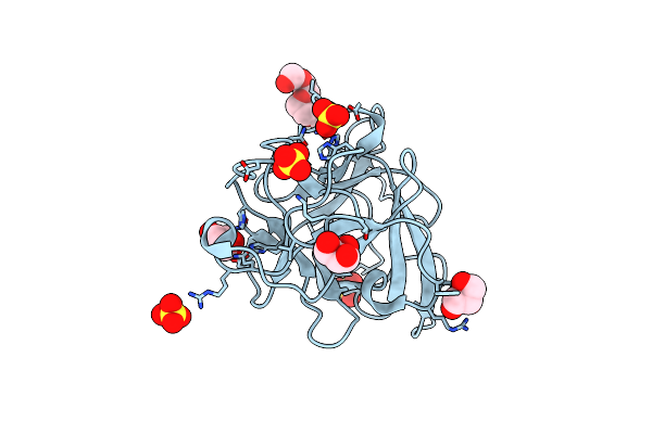 |
Organism: Saccharomyces cerevisiae
Method: X-RAY DIFFRACTION Resolution:1.60 Å Release Date: 2020-12-23 Classification: PEPTIDE BINDING PROTEIN Ligands: PG4, GOL, SO4 |
 |
Organism: Saccharomyces cerevisiae
Method: X-RAY DIFFRACTION Resolution:1.90 Å Release Date: 2020-12-23 Classification: PEPTIDE BINDING PROTEIN Ligands: GOL |
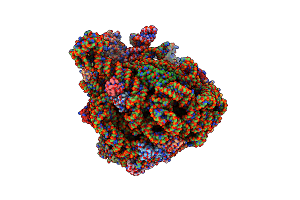 |
Cryo-Em Structure Of The 50S Ribosomal Subunit At 2.58 Angstroms With Modeled Gbc Secm Peptide
Organism: Bos taurus, Escherichia coli bl21(de3)
Method: ELECTRON MICROSCOPY Release Date: 2020-09-30 Classification: RIBOSOME |