
Search Count: 34
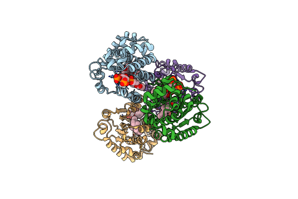 |
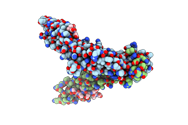
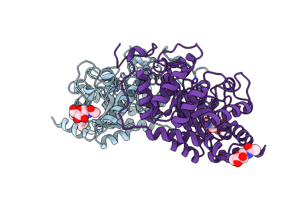
Crystal Structure Of Mouse Alpha-Tocopherol Transfer Protein In Complex With Alpha-Tocopherol And Phosphatidylinositol-(3,4)-Bisphosphate
Organism: Mus musculus
Method: X-RAY DIFFRACTION Resolution:2.61 Å Release Date: 2013-05-01 Classification: TRANSPORT PROTEIN Ligands: VIV, 3PT |
 |
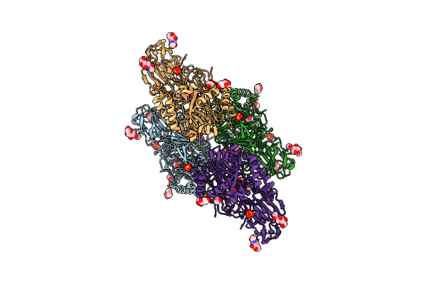
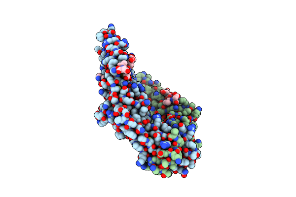
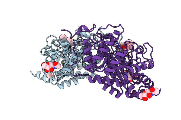
Crystal Structure Of Mouse Alpha-Tocopherol Transfer Protein In Complex With Alpha-Tocopherol And Phosphatidylinositol-(4,5)-Bisphosphate
Organism: Mus musculus
Method: X-RAY DIFFRACTION Resolution:2.05 Å Release Date: 2013-05-01 Classification: TRANSPORT PROTEIN Ligands: VIV, 4PT, PBU, PO4 |
 |
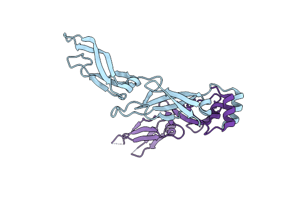
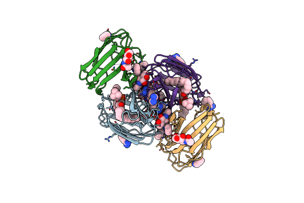
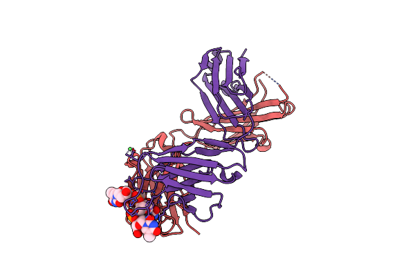
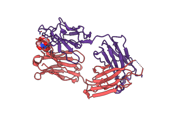
Antibody 64M-5 Fab In Complex With A Double-Stranded Dna (6-4) Photoproduct
Organism: Mus musculus
Method: X-RAY DIFFRACTION Resolution:2.50 Å Release Date: 2013-03-27 Classification: IMMUNE SYSTEM/DNA Ligands: NCO |
 |
Organism: Homo sapiens
Method: X-RAY DIFFRACTION Resolution:1.80 Å Release Date: 2011-12-07 Classification: HYDROLASE Ligands: NAG, CL, GAL, SO4, EDO |
 |
Crystal Structure Of Human Beta-Galactosidase In Complex With 1-Deoxygalactonojirimycin
Organism: Homo sapiens
Method: X-RAY DIFFRACTION Resolution:1.79 Å Release Date: 2011-12-07 Classification: HYDROLASE Ligands: NAG, CL, DGJ, SO4, EDO |
 |
Organism: Homo sapiens
Method: X-RAY DIFFRACTION Resolution:1.85 Å Release Date: 2011-02-23 Classification: CHAPERONE |
 |
Crystal Structure Of Human Hsp40 Hdj1 Peptide-Binding Domain Complexed With A C-Terminal Peptide Of Hsp70
Organism: Homo sapiens
Method: X-RAY DIFFRACTION Resolution:1.85 Å Release Date: 2011-02-23 Classification: CHAPERONE |
 |
Crystal Structure Of Human Hsp40 Hdj1 Peptide-Binding Domain Complexed With A C-Terminal Peptide Of Hsp70
Organism: Homo sapiens
Method: X-RAY DIFFRACTION Resolution:2.51 Å Release Date: 2011-02-23 Classification: CHAPERONE |
 |
Crystal Structure Of The Sep22 Dodecamer, A Dps-Like Protein From Salmonella Enterica Subsp. Enterica Serovar Enteritidis
Organism: Salmonella enterica subsp. enterica serovar kentucky
Method: X-RAY DIFFRACTION Resolution:1.25 Å Release Date: 2011-01-12 Classification: METAL BINDING PROTEIN, Oxidoreductase Ligands: SO4, MG |
 |
Crystal Structure Of The Sep22 Dodecamer, A Dps-Like Protein From Salmonella Enterica Subsp. Enterica Serovar Enteritidis, Fe-Soaked Form
Organism: Salmonella enterica subsp. enterica serovar kentucky
Method: X-RAY DIFFRACTION Resolution:1.30 Å Release Date: 2011-01-12 Classification: METAL BINDING PROTEIN, Oxidoreductase Ligands: FE2, SO4, MG |
 |
Organism: Mus musculus
Method: X-RAY DIFFRACTION Resolution:1.65 Å Release Date: 2010-06-09 Classification: IMMUNE SYSTEM Ligands: NAG, L9R, BEN |
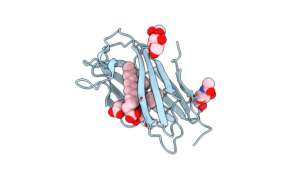 |
Organism: Homo sapiens
Method: X-RAY DIFFRACTION Resolution:2.00 Å Release Date: 2007-06-26 Classification: LIPID BINDING PROTEIN Ligands: NAG, MYR |
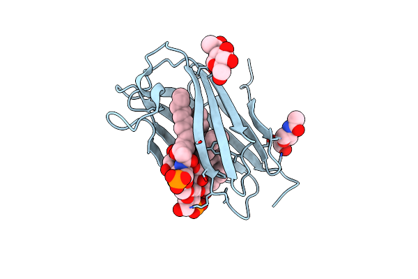 |
Organism: Homo sapiens
Method: X-RAY DIFFRACTION Resolution:2.21 Å Release Date: 2007-06-26 Classification: LIPID BINDING PROTEIN Ligands: NAG, LP5, LP4 |
 |
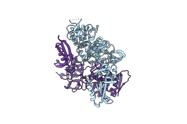
Crystal Structure Of The Vma1-Derived Endonuclease With The Ligated Extein Segment
Organism: Saccharomyces cerevisiae
Method: X-RAY DIFFRACTION Resolution:2.90 Å Release Date: 2004-09-22 Classification: HYDROLASE |
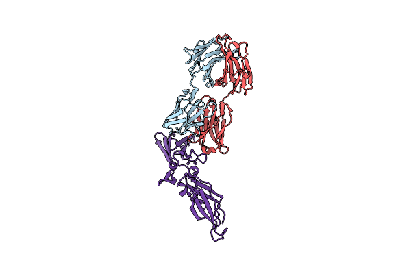 |
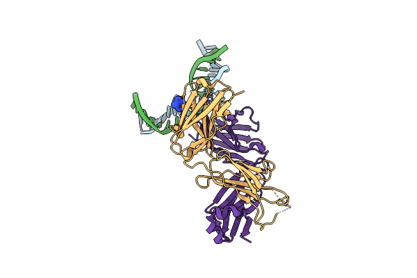
Crystal Structure Of A Humanized Fab Fragment Of Anti-Tissue-Factor Antibody In Complex With Tissue Factor
Organism: Homo sapiens
Method: X-RAY DIFFRACTION Resolution:2.10 Å Release Date: 2004-07-25 Classification: IMMUNE SYSTEM/BLOOD CLOTTING |
 |
Organism: Mus musculus
Method: X-RAY DIFFRACTION Resolution:2.40 Å Release Date: 2002-11-15 Classification: IMMUNE SYSTEM/DNA Ligands: NI |
 |
Crystal Structure Of The Vma1-Derived Endonuclease Bearing The N And C Extein Propeptides
Organism: Saccharomyces cerevisiae
Method: X-RAY DIFFRACTION Resolution:2.10 Å Release Date: 2002-08-29 Classification: HYDROLASE |
 |
Organism: Homo sapiens
Method: X-RAY DIFFRACTION Resolution:2.30 Å Release Date: 2002-08-28 Classification: HYDROLASE Ligands: NAG, ZN |
 |
Organism: Homo sapiens
Method: X-RAY DIFFRACTION Resolution:2.00 Å Release Date: 2002-08-28 Classification: HYDROLASE Ligands: NAG, ZN, CIL |
 |
Organism: Mus musculus
Method: X-RAY DIFFRACTION Resolution:2.40 Å Release Date: 2001-02-21 Classification: IMMUNE SYSTEM |