
Search Count: 37
 |
Organism: Escherichia coli
Method: X-RAY DIFFRACTION Resolution:1.90 Å Release Date: 2024-12-11 Classification: DNA BINDING PROTEIN Ligands: ZN, FE2, EPE, SO4, NI, CL |
 |
Organism: Staphylococcus aureus
Method: X-RAY DIFFRACTION Resolution:1.90 Å Release Date: 2024-07-10 Classification: HYDROLASE Ligands: FE, ZN, PO4, CL |
 |
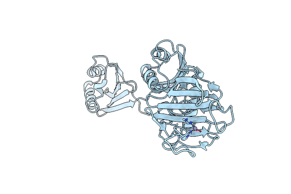
Organism: Staphylococcus aureus
Method: X-RAY DIFFRACTION Resolution:1.05 Å Release Date: 2023-09-13 Classification: HYDROLASE Ligands: FE, ZN, SO4 |
 |
Organism: Staphylococcus aureus
Method: X-RAY DIFFRACTION Resolution:1.50 Å Release Date: 2023-09-06 Classification: HYDROLASE Ligands: FE, ZN, PO4, CL |
 |
Organism: Vibrio cholerae
Method: X-RAY DIFFRACTION Resolution:2.30 Å Release Date: 2020-05-27 Classification: HYDROLASE Ligands: ZN |
 |
Organism: Vibrio cholerae serotype o1 (strain atcc 39315 / el tor inaba n16961)
Method: X-RAY DIFFRACTION Resolution:2.08 Å Release Date: 2020-05-27 Classification: HYDROLASE Ligands: ZN, GOL |
 |
Alanine-Glyoxylate Aminotransferase 1 (Agt1) From Arabidopsis Thaliana In Presence Of Serine
Organism: Arabidopsis thaliana
Method: X-RAY DIFFRACTION Resolution:2.10 Å Release Date: 2019-10-23 Classification: TRANSFERASE Ligands: PLP, 3PY |
 |
Organism: Arabidopsis thaliana
Method: X-RAY DIFFRACTION Resolution:2.18 Å Release Date: 2019-10-23 Classification: TRANSFERASE Ligands: PLP, FMT, CL |
 |
Organism: Escherichia coli (strain k12)
Method: X-RAY DIFFRACTION Resolution:2.10 Å Release Date: 2019-05-08 Classification: BIOSYNTHETIC PROTEIN |
 |
Lpoa N-Terminal Domain From Haemophilus Influenzae; Monoclinic Form At 1.35 A Resolution
Organism: Haemophilus influenzae
Method: X-RAY DIFFRACTION Resolution:1.35 Å Release Date: 2019-05-01 Classification: BIOSYNTHETIC PROTEIN Ligands: CL |
 |
Crystal Structure Of Full-Length Lpoa From Haemophilus Influenzae At 1.97 Angstrom Resolution
Organism: Haemophilus influenzae
Method: X-RAY DIFFRACTION Resolution:1.97 Å Release Date: 2017-09-13 Classification: BIOSYNTHETIC PROTEIN Ligands: CL |
 |
Haemophilus Influenzae Lpoa: Monoclinic Form (Mon2) With 2 Molecules Per A.U.
Organism: Haemophilus influenzae (strain atcc 51907 / dsm 11121 / kw20 / rd)
Method: X-RAY DIFFRACTION Resolution:2.60 Å Release Date: 2017-09-13 Classification: BIOSYNTHETIC PROTEIN |
 |
Crystal Structure Of Full-Length Lpoa, Monoclinic Form 1, From Haemophilus Influenzae
Organism: Haemophilus influenzae (strain atcc 51907 / dsm 11121 / kw20 / rd)
Method: X-RAY DIFFRACTION Resolution:2.80 Å Release Date: 2017-09-13 Classification: BIOSYNTHETIC PROTEIN Ligands: CL |
 |
Crystal Structure Of The Lpoa N-Terminal Domain From Haemophilus Influenzae
Organism: Haemophilus influenzae
Method: X-RAY DIFFRACTION Resolution:1.95 Å Release Date: 2015-03-04 Classification: PROTEIN BINDING Ligands: SO4, GOL, CL |
 |
Structure Of Gfcc (Ymcb), Protein Encoded By The E. Coli Group 4 Capsule Operon
Organism: Escherichia coli o127:h6
Method: X-RAY DIFFRACTION Resolution:1.91 Å Release Date: 2011-04-06 Classification: UNKNOWN FUNCTION Ligands: SO4 |
 |
Organism: Haemophilus influenzae
Method: X-RAY DIFFRACTION Resolution:1.35 Å Release Date: 2008-05-13 Classification: BIOSYNTHETIC PROTEIN Ligands: SO4, BME |
 |
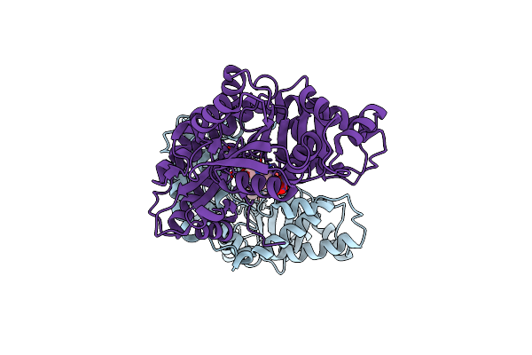
Crystal Structure Of Yersinia Protein Tyrosine Phosphatase Complexed With Vanadate, A Transition State Analogue
Organism: Yersinia enterocolitica
Method: X-RAY DIFFRACTION Resolution:2.20 Å Release Date: 2006-09-19 Classification: HYDROLASE Ligands: VO4 |
 |
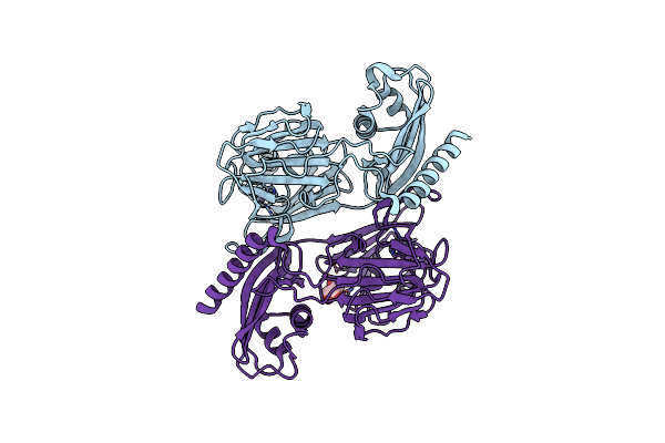
Yersinia Yoph (Residues 163-468) C403S Binds Phosphotyrosyl Peptide At Two Sites
Organism: Yersinia enterocolitica
Method: X-RAY DIFFRACTION Resolution:3.00 Å Release Date: 2005-03-22 Classification: HYDROLASE/HYDROLASE INHIBITOR |
 |
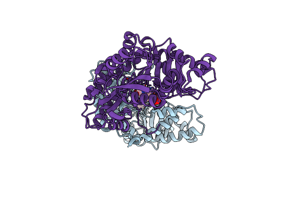
Yersinia Yoph (Residues 163-468) Binds Phosphonodifluoromethyl-Phe Containing Hexapeptide At Two Sites
Organism: Yersinia enterocolitica
Method: X-RAY DIFFRACTION Resolution:2.50 Å Release Date: 2005-03-22 Classification: HYDROLASE |
 |
Nmr Structure Of The Type Iii Secretory Domain Of Yersinia Yoph Complexed With The Skap-Hom Phospho-Peptide N-Acetyl-Depyddpf-Nh2
Organism: Yersinia pseudotuberculosis
Method: SOLUTION NMR Release Date: 2002-07-24 Classification: HYDROLASE |