
Search Count: 34
 |
Organism: Stichodactyla helianthus
Method: ELECTRON MICROSCOPY Release Date: 2025-10-29 Classification: MEMBRANE PROTEIN Ligands: FO4, CLR |
 |
Organism: Actinia fragacea
Method: ELECTRON MICROSCOPY Release Date: 2025-10-29 Classification: MEMBRANE PROTEIN Ligands: FO4, CLR |
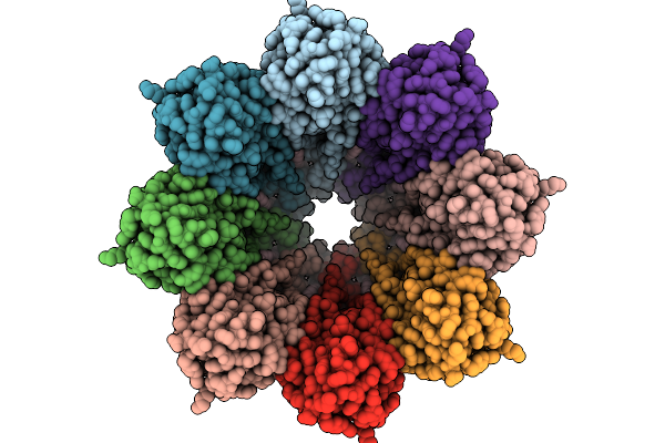 |
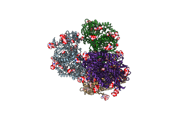
Structure Of The Octameric Pore Of Fragaceotxin C (Frac Or Delta-Actitoxin-Afr1A) In Large Unilamellar Vesicles.
Organism: Actinia fragacea
Method: ELECTRON MICROSCOPY Release Date: 2025-10-08 Classification: MEMBRANE PROTEIN Ligands: FO4, CLR |
 |
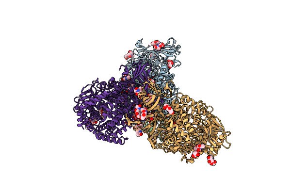
Structure Of 6Mer Pore Intermediate Of Sticholysin Ii (Stnii) Toxin In Lipid Nanodiscs
Organism: Stichodactyla helianthus
Method: ELECTRON MICROSCOPY Release Date: 2025-10-08 Classification: MEMBRANE PROTEIN Ligands: FO4 |
 |
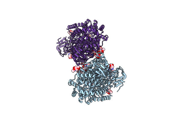
Structure Of 5Mer Pore Intermediate Of Sticholysin Ii (Stnii) Toxin In Lipid Nanodiscs
Organism: Stichodactyla helianthus
Method: ELECTRON MICROSCOPY Release Date: 2025-09-17 Classification: MEMBRANE PROTEIN Ligands: FO4 |
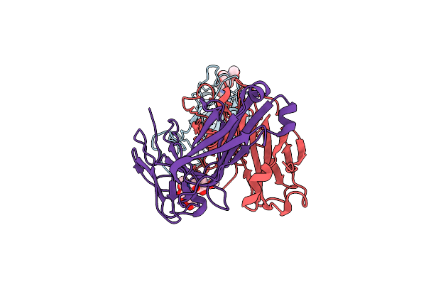 |
Sars-Cov-2 Spike In Complex With The 17T2 Neutralizing Antibody Fab Fragment (Local Refinement Of Rbd And Fab)
Organism: Severe acute respiratory syndrome coronavirus, Homo sapiens
Method: ELECTRON MICROSCOPY Release Date: 2024-01-10 Classification: VIRAL PROTEIN |
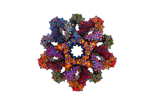 |
3D Reconstruction Of The Cylindrical Assembly Of Dnaja2 Delta G/F By Imposing D5 Symmetry
Organism: Saccharomyces cerevisiae s288c, Homo sapiens
Method: ELECTRON MICROSCOPY Resolution:6.90 Å Release Date: 2023-07-26 Classification: CHAPERONE Ligands: ZN |
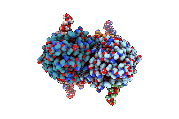 |
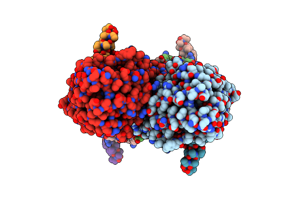
Partial Structure Of Tyrosine Hydroxylase Lacking The First 35 Residues In Complex With Dopamine.
Organism: Homo sapiens
Method: ELECTRON MICROSCOPY Release Date: 2021-12-22 Classification: OXIDOREDUCTASE Ligands: LDP, FE |
 |
Partial Structure Of Tyrosine Hydroxylase In Complex With Dopamine Showing The Catalytic Domain And An Alpha-Helix From The Regulatory Domain Involved In Dopamine Binding.
Organism: Homo sapiens
Method: ELECTRON MICROSCOPY Release Date: 2021-12-08 Classification: OXIDOREDUCTASE Ligands: LDP, FE |
 |
Organism: Homo sapiens
Method: ELECTRON MICROSCOPY Release Date: 2021-12-01 Classification: OXIDOREDUCTASE Ligands: FE |
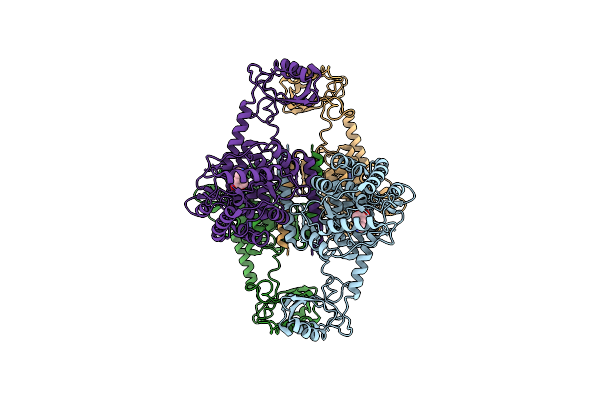 |
Atomic Model Of The Em-Based Structure Of The Full-Length Tyrosine Hydroxylase In Complex With Dopamine (Residues 40-497) In Which The Regulatory Domain (Residues 40-165) Has Been Included Only With The Backbone Atoms
Organism: Homo sapiens
Method: ELECTRON MICROSCOPY Release Date: 2021-11-17 Classification: OXIDOREDUCTASE Ligands: FE, LDP |
 |
Organism: Homo sapiens
Method: ELECTRON MICROSCOPY Release Date: 2021-11-17 Classification: OXIDOREDUCTASE Ligands: FE |
 |
Organism: Homo sapiens
Method: ELECTRON MICROSCOPY Release Date: 2019-07-03 Classification: CHAPERONE Ligands: ADP |
 |
Organism: Pseudomonas syringae
Method: X-RAY DIFFRACTION Resolution:1.10 Å Release Date: 2019-04-03 Classification: PROTEIN FIBRIL Ligands: HOH |
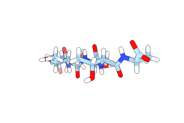 |
Organism: Pseudomonas syringae
Method: X-RAY DIFFRACTION Resolution:1.10 Å Release Date: 2019-04-03 Classification: PROTEIN FIBRIL |
 |
Organism: Pseudomonas syringae pv. syringae
Method: ELECTRON CRYSTALLOGRAPHY Resolution:0.90 Å Release Date: 2019-03-27 Classification: PROTEIN FIBRIL Ligands: HOH |
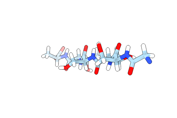 |
Organism: Pseudomonas syringae pv. syringae
Method: ELECTRON CRYSTALLOGRAPHY Resolution:0.90 Å Release Date: 2019-03-27 Classification: PROTEIN FIBRIL |
 |
Organism: Sus scrofa
Method: X-RAY DIFFRACTION Resolution:2.50 Å Release Date: 2017-04-12 Classification: HYDROLASE Ligands: NAG, ZN |
 |
Organism: Sus scrofa
Method: X-RAY DIFFRACTION Resolution:2.00 Å Release Date: 2017-04-05 Classification: HYDROLASE Ligands: ZN, NAG, ACT |
 |
Organism: Homo sapiens
Method: X-RAY DIFFRACTION Resolution:2.60 Å Release Date: 2017-04-05 Classification: HYDROLASE Ligands: NAG, ZN, EDO |