
Search Count: 37
 |
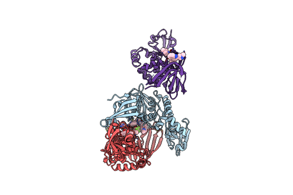
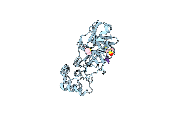
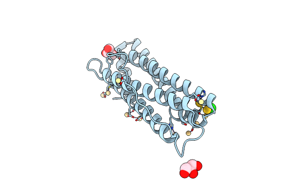
Organism: Homo sapiens
Method: X-RAY DIFFRACTION Resolution:2.16 Å Release Date: 2024-11-27 Classification: TRANSFERASE/TRANSFERASE INHIBITOR Ligands: ADP, MG, K, A1A40, PO4 |
 |
Organism: Severe acute respiratory syndrome coronavirus 2
Method: X-RAY DIFFRACTION Resolution:2.60 Å Release Date: 2024-10-02 Classification: VIRAL PROTEIN Ligands: A1AZ1, TFA, ZN |
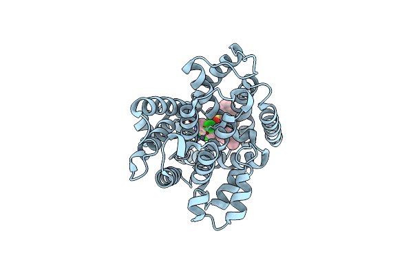 |
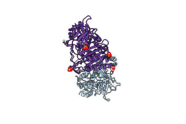
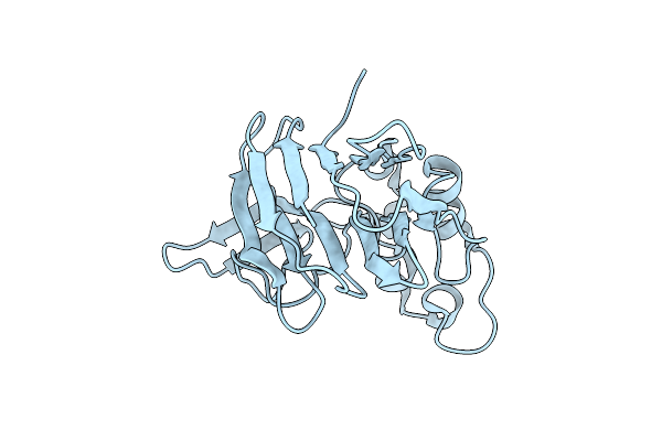
Organism: Homo sapiens
Method: X-RAY DIFFRACTION Resolution:1.89 Å Release Date: 2023-04-26 Classification: HYDROLASE Ligands: ZN, MG, VL9 |
 |
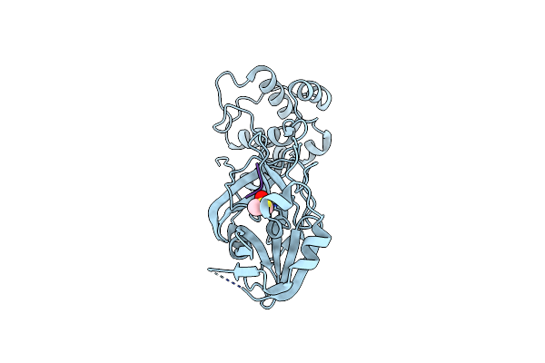
Organism: Porphyridium purpureum
Method: X-RAY DIFFRACTION Resolution:1.60 Å Release Date: 2023-02-08 Classification: PHOTOSYNTHESIS Ligands: PEB, SO4 |
 |
Crystal Structure Of Sars Cov-2 Main Protease In Complex With An Inhibitor 57
Organism: Severe acute respiratory syndrome coronavirus 2, Synthetic construct
Method: X-RAY DIFFRACTION Resolution:1.34 Å Release Date: 2022-12-07 Classification: ANTIVIRAL PROTEIN Ligands: DMS |
 |
Crystal Structure Of Sars Cov-2 Main Protease In Complex With An Inhibitor 58
Organism: Severe acute respiratory syndrome coronavirus 2, Synthetic construct
Method: X-RAY DIFFRACTION Resolution:1.94 Å Release Date: 2022-12-07 Classification: ANTIVIRAL PROTEIN Ligands: DMS, OCA |
 |
Crystal Structure Of Sars Cov-2 Main Protease In Complex With An Inhibitor 4
Organism: Severe acute respiratory syndrome coronavirus 2, Synthetic construct
Method: X-RAY DIFFRACTION Resolution:2.00 Å Release Date: 2022-11-30 Classification: ANTIVIRAL PROTEIN Ligands: DMS |
 |
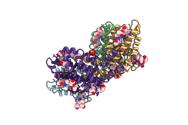
Organism: Homo sapiens
Method: X-RAY DIFFRACTION Resolution:1.90 Å Release Date: 2020-08-26 Classification: METAL BINDING PROTEIN Ligands: EF2, ZN |
 |
Organism: Homo sapiens
Method: X-RAY DIFFRACTION Resolution:1.80 Å Release Date: 2020-08-26 Classification: METAL BINDING PROTEIN Ligands: F4U, ZN, SO4 |
 |
Organism: Streptomyces albidoflavus
Method: X-RAY DIFFRACTION Resolution:2.20 Å Release Date: 2020-06-24 Classification: HYDROLASE Ligands: SO4, MLI, YT3 |
 |
Organism: Streptomyces albidoflavus
Method: X-RAY DIFFRACTION Resolution:2.42 Å Release Date: 2020-06-24 Classification: HYDROLASE Ligands: DLE, SO4, YT3, E7L |
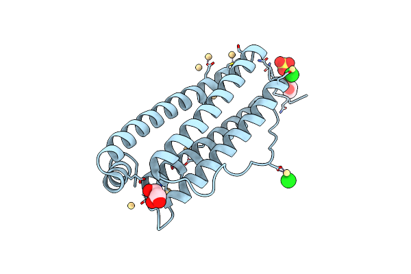 |
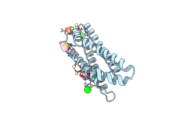
Organism: Equus caballus
Method: X-RAY DIFFRACTION Resolution:1.43 Å Release Date: 2019-02-06 Classification: METAL TRANSPORT Ligands: CD, CL, SO4, GOL |
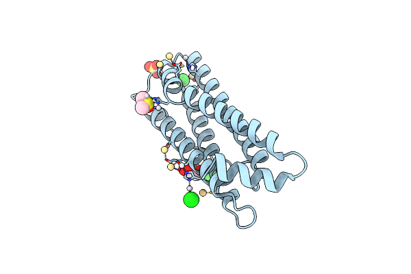 |
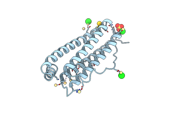
The X-Ray Structure Of The Horse Spleen Ferritin Nanocage Containing Pt, Obtained Upon Encapsulation Of A Pt(Ii) Terpyridine Compound Within The Protein Cage
Organism: Equus caballus
Method: X-RAY DIFFRACTION Resolution:1.33 Å Release Date: 2018-12-19 Classification: METAL TRANSPORT Ligands: CD, CL, PT, SO4, GOL, DMS |
 |
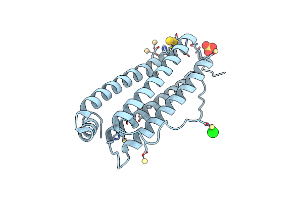
The X-Ray Structure Of The Horse Spleen Ferritin Nanocage Containing Pt, Obtained Upon Encapsulation Of A Pt(Ii) Terpyridine Compound Within The Protein Cage
Organism: Equus caballus
Method: X-RAY DIFFRACTION Resolution:1.58 Å Release Date: 2018-12-19 Classification: METAL TRANSPORT Ligands: CD, CL, PT, SO4, GOL, DMS |
 |
Crystal Structure Of Alginate Lyase From Flavobacterium Sp. Umi-01 Reveals Polymannuronate Specificity
Organism: Flavobacterium sp. umi-01
Method: X-RAY DIFFRACTION Resolution:1.54 Å Release Date: 2018-07-04 Classification: STRUCTURAL PROTEIN |
 |
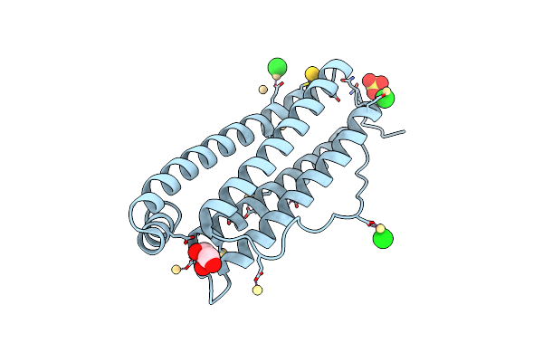
The X-Ray Structure Of The Ferritin Nanocage Containing Au And Pt, Obtained Upon Encapsulation Of A Single Heterobimetallic Compound Within The Protein Cage (Rotating Anode Data)
Organism: Equus caballus
Method: X-RAY DIFFRACTION Resolution:1.80 Å Release Date: 2018-05-16 Classification: METAL TRANSPORT Ligands: CD, CL, SO4, AU |
 |
The X-Ray Structure Of The Ferritin Nanocage Containing Au And Pt, Obtained Upon Encapsulation Of A Single Heterobimetallic Compound Within The Protein Cage (Synchrtron Data)
Organism: Equus caballus
Method: X-RAY DIFFRACTION Resolution:1.50 Å Release Date: 2018-05-16 Classification: METAL TRANSPORT Ligands: CD, CL, SO4, GOL, AU |
 |
Organism: Equus caballus
Method: X-RAY DIFFRACTION Resolution:1.82 Å Release Date: 2017-12-13 Classification: PROTEIN TRANSPORT Ligands: CD, CL, SO4, AU |
 |
Organism: Equus caballus
Method: X-RAY DIFFRACTION Resolution:2.60 Å Release Date: 2017-12-13 Classification: PROTEIN TRANSPORT Ligands: CD, CL, GOL, AU |
 |
Organism: Thaumatococcus daniellii
Method: X-RAY DIFFRACTION Resolution:1.30 Å Release Date: 2017-08-09 Classification: PLANT PROTEIN |