
Search Count: 49
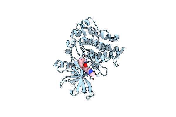 |
Organism: Homo sapiens
Method: X-RAY DIFFRACTION Release Date: 2025-06-25 Classification: CELL CYCLE Ligands: A1A6M, CL, MG |
 |
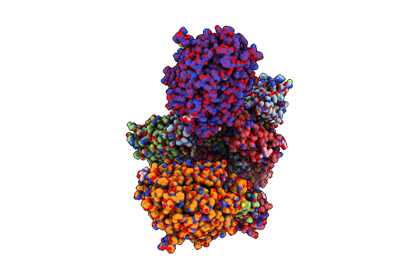
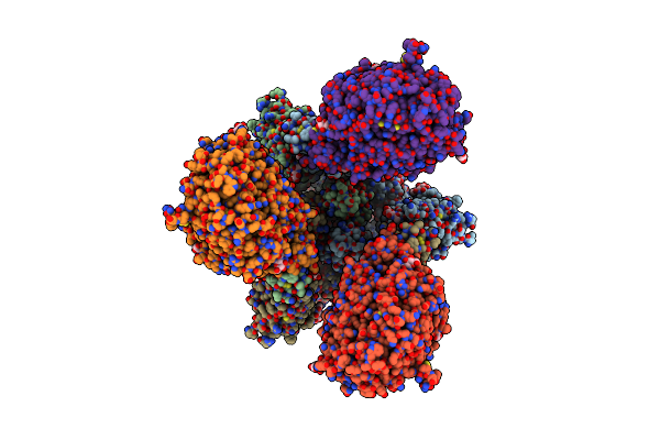
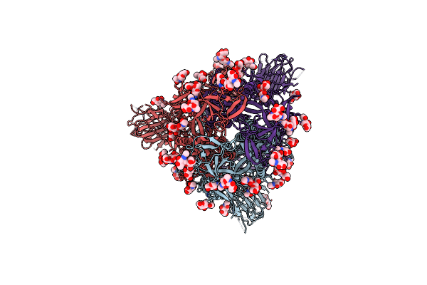
Organism: Severe acute respiratory syndrome coronavirus 2, Homo sapiens
Method: ELECTRON MICROSCOPY Release Date: 2020-12-16 Classification: HYDROLASE/VIRAL PROTEIN Ligands: NAG |
 |
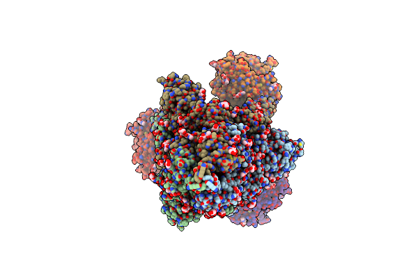
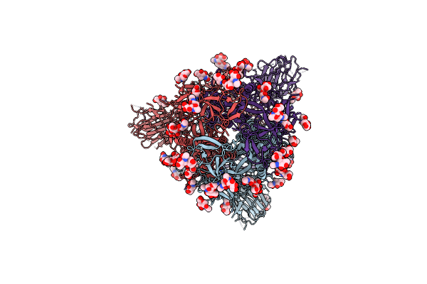
Organism: Severe acute respiratory syndrome coronavirus 2, Homo sapiens
Method: ELECTRON MICROSCOPY Release Date: 2020-12-16 Classification: HYDROLASE/VIRAL PROTEIN Ligands: NAG |
 |
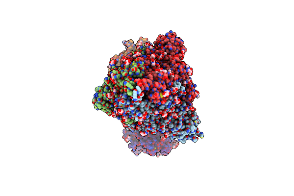
Organism: Severe acute respiratory syndrome coronavirus 2, Homo sapiens
Method: ELECTRON MICROSCOPY Release Date: 2020-12-16 Classification: VIRAL PROTEIN/Hydrolase Ligands: NAG |
 |
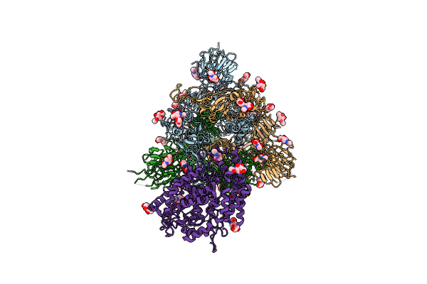
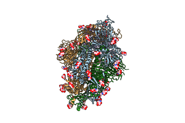
Ace2-Rbd Focused Refinement Using Symmetry Expansion Of Applied C3 For Triple Ace2-Bound Sars-Cov-2 Trimer Spike At Ph 7.4
Organism: Homo sapiens, Severe acute respiratory syndrome coronavirus 2
Method: ELECTRON MICROSCOPY Release Date: 2020-12-09 Classification: Hydrolase/Viral Protein Ligands: NAG |
 |
Organism: Homo sapiens, Severe acute respiratory syndrome coronavirus 2
Method: ELECTRON MICROSCOPY Release Date: 2020-12-09 Classification: HYDROLASE/VIRAL PROTEIN Ligands: NAG |
 |
Organism: Severe acute respiratory syndrome coronavirus 2, Homo sapiens
Method: ELECTRON MICROSCOPY Release Date: 2020-12-09 Classification: HYDROLASE/VIRAL PROTEIN Ligands: NAG |
 |
Organism: Severe acute respiratory syndrome coronavirus 2, Homo sapiens
Method: ELECTRON MICROSCOPY Release Date: 2020-12-09 Classification: HYDROLASE/VIRAL PROTEIN Ligands: NAG |
 |
Organism: Severe acute respiratory syndrome coronavirus 2
Method: ELECTRON MICROSCOPY Release Date: 2020-11-25 Classification: VIRAL PROTEIN Ligands: NAG |
 |
Organism: Severe acute respiratory syndrome coronavirus 2
Method: ELECTRON MICROSCOPY Release Date: 2020-08-12 Classification: VIRAL PROTEIN Ligands: NAG |
 |
Organism: Severe acute respiratory syndrome coronavirus 2
Method: ELECTRON MICROSCOPY Release Date: 2020-08-12 Classification: VIRAL PROTEIN Ligands: NAG |
 |
Organism: Severe acute respiratory syndrome coronavirus 2
Method: ELECTRON MICROSCOPY Release Date: 2020-08-12 Classification: VIRAL PROTEIN Ligands: NAG |
 |
Organism: Severe acute respiratory syndrome coronavirus 2
Method: ELECTRON MICROSCOPY Release Date: 2020-08-12 Classification: VIRAL PROTEIN Ligands: NAG |
 |
Organism: Severe acute respiratory syndrome coronavirus 2
Method: ELECTRON MICROSCOPY Release Date: 2020-07-29 Classification: VIRAL PROTEIN Ligands: NAG |
 |
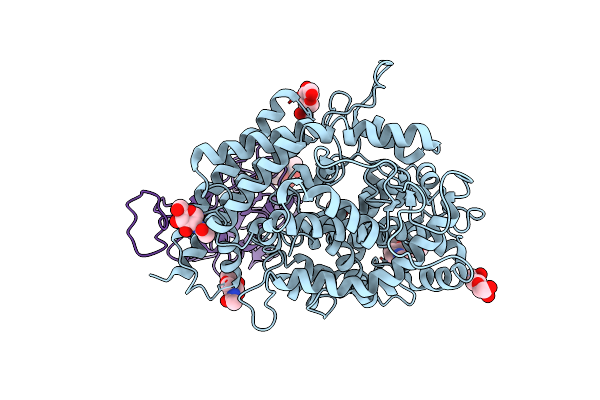
Structure Of A Modified Protein Containing A Genetically Encoded Phosphoserine
Organism: Homo sapiens
Method: X-RAY DIFFRACTION Resolution:1.66 Å Release Date: 2018-10-24 Classification: CHAPERONE Ligands: ADP, PO4, MG, K |
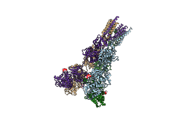 |
Organism: Rattus norvegicus
Method: X-RAY DIFFRACTION Resolution:4.00 Å Release Date: 2016-10-19 Classification: MEMBRANE PROTEIN, TRANSPORT PROTEIN Ligands: NAG |
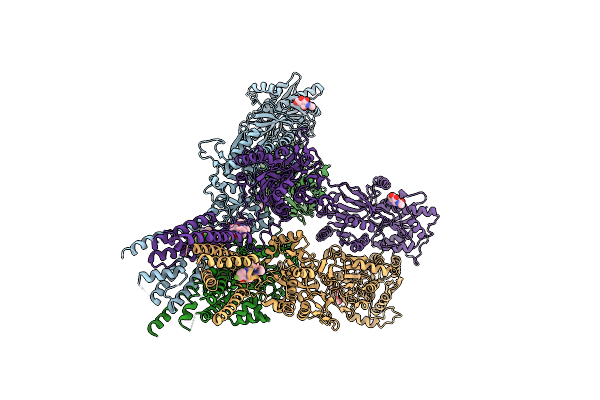 |
Ampa Subtype Ionotropic Glutamate Receptor Glua2 In Complex With Noncompetitive Inhibitor Cp465022
Organism: Rattus norvegicus
Method: X-RAY DIFFRACTION Resolution:4.37 Å Release Date: 2016-10-19 Classification: TRANSPORT PROTEIN/INHIBITOR Ligands: NAG, 6ZQ |
 |
Ampa Subtype Ionotropic Glutamate Receptor Glua2 In Complex With Noncompetitive Inhibitor Perampanel
Organism: Rattus norvegicus
Method: X-RAY DIFFRACTION Resolution:4.00 Å Release Date: 2016-10-19 Classification: TRANSPORT PROTEIN/INHIBITOR Ligands: NAG, 6ZP |
 |
Organism: Rattus norvegicus
Method: X-RAY DIFFRACTION Resolution:4.51 Å Release Date: 2016-10-19 Classification: TRANSPORT PROTEIN/INHIBITOR Ligands: NAG, GYB |
 |
Ampa Subtype Ionotropic Glutamate Receptor Glua2 In Complex With Noncompetitive Inhibitor Gyki53655
Organism: Rattus norvegicus
Method: X-RAY DIFFRACTION Resolution:3.80 Å Release Date: 2016-10-19 Classification: TRANSPORT PROTEIN/INHIBITOR Ligands: NAG, GYK |