
Search Count: 15
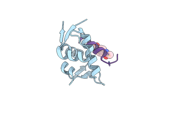 |
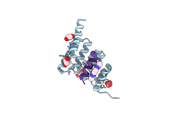
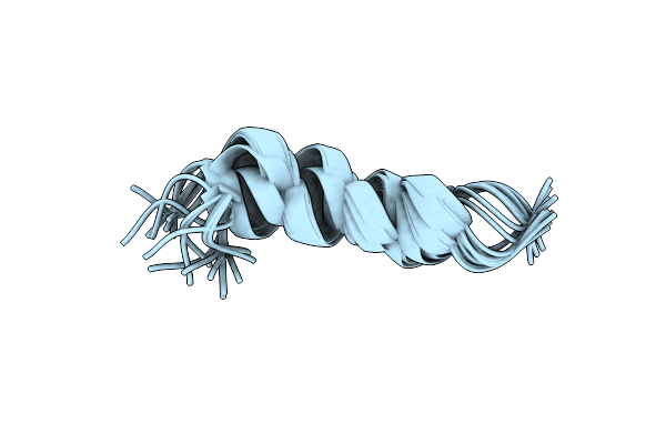
Structure Of The Mdm2 P53 Binding Domain In Complex With H101, An All-D Helicon Polypeptide
Organism: Homo sapiens, Synthetic construct
Method: X-RAY DIFFRACTION Resolution:1.61 Å Release Date: 2023-02-15 Classification: LIGASE Ligands: CL, WHL |
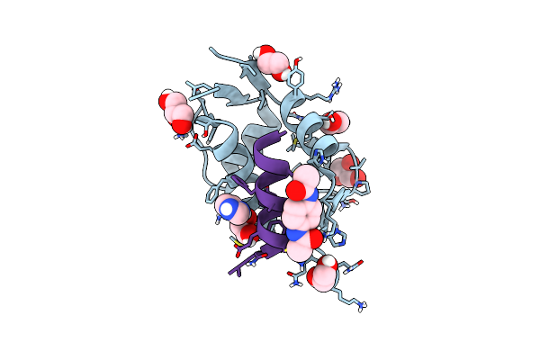 |
Structure Of The Mdm2 P53 Binding Domain In Complex With H102, An All-D Helicon Polypeptide
Organism: Homo sapiens, Synthetic construct
Method: X-RAY DIFFRACTION Resolution:1.28 Å Release Date: 2023-02-15 Classification: LIGASE Ligands: EDO, CL, GOL, WHL, IMD |
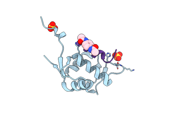 |
Structure Of The Mdm2 P53 Binding Domain In Complex With H103, An All-D Helicon Polypeptide
Organism: Homo sapiens, Synthetic construct
Method: X-RAY DIFFRACTION Resolution:1.86 Å Release Date: 2023-02-15 Classification: LIGASE Ligands: SO4, CL, WHL |
 |
Structure Of The Mdm2 P53 Binding Domain In Complex With H103, An All-D Helicon Polypeptide, Alternative C-Terminus
Organism: Homo sapiens, Synthetic construct
Method: X-RAY DIFFRACTION Resolution:1.40 Å Release Date: 2023-02-15 Classification: LIGASE Ligands: EDO, SO4, DMS, WHL |
 |
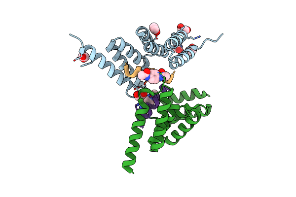
Structure Of The Stub1 Tpr Domain In Complex With H201, An All-D Helicon Polypeptide
Organism: Homo sapiens, Synthetic construct
Method: X-RAY DIFFRACTION Resolution:1.69 Å Release Date: 2023-02-15 Classification: LIGASE Ligands: SO4, EDO, WHL |
 |
Structure Of The Stub1 Tpr Domain In Complex With H202, An All-D Helicon Polypeptide
Organism: Homo sapiens, Synthetic construct
Method: X-RAY DIFFRACTION Resolution:1.73 Å Release Date: 2023-02-15 Classification: LIGASE Ligands: EDO, WHL |
 |
Structure Of The Stub1 Tpr Domain In Complex With H203, An All-D Helicon Polypeptide
Organism: Homo sapiens, Synthetic construct
Method: X-RAY DIFFRACTION Resolution:1.56 Å Release Date: 2023-02-15 Classification: LIGASE Ligands: EDO, WHL |
 |
Structure Of The Stub1 Tpr Domain In Complex With H204, An All-D Helicon Polypeptide
Organism: Homo sapiens, Synthetic construct
Method: X-RAY DIFFRACTION Resolution:2.21 Å Release Date: 2023-02-15 Classification: LIGASE Ligands: WHL, EDO |
 |
Changing Abra Protein Peptide To Fit The Hla-Dr B1*0301 Molecule Renders It Protection-Inducing
Organism: Plasmodium falciparum
Method: SOLUTION NMR Release Date: 2015-09-16 Classification: PEPTIDE BINDING PROTEIN |
 |
Organism: Mus musculus
Method: X-RAY DIFFRACTION Resolution:2.50 Å Release Date: 2015-04-01 Classification: IMMUNE SYSTEM |
 |
Evidence Supporting The Hypothesis That Specifically Modifying A Malaria Peptide To Fit Into Hla-Dr 1*03 Molecules Induces Antibody Production And Protection
Organism: Plasmodium falciparum camp/malaysia
Method: SOLUTION NMR Release Date: 2015-02-04 Classification: PROTEIN BINDING |
 |
Protection Against Experimental P. Falciparum Malaria Is Associated With Short Ama-1 Peptide Analogue Alpha-Helical Structures
Organism: Plasmodium falciparum
Method: SOLUTION NMR Release Date: 2015-02-04 Classification: PROTEIN BINDING |
 |
Structure Immunogenicity And Protectivity Relationship For The 1585 Malarial Peptide And Its Substitution Analogues
Organism: Plasmodium falciparum
Method: SOLUTION NMR Release Date: 2014-10-08 Classification: CELL INVASION |
 |
Protective Cellular Immunity Against P. Falciparum Malaria Merozoite Is Associated With A Different P7 And P8 Residue Orientation In The Mhc-Peptide-Tcr Complex
Organism: Plasmodium falciparum
Method: SOLUTION NMR Release Date: 2014-10-01 Classification: PEPTIDE BINDING PROTEIN |
 |
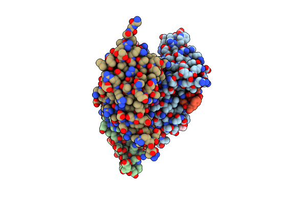
Tarantula Heavy Meromyosin Obtained By Flexible Docking To Tarantula Muscle Thick Filament Cryo-Em 3D-Map
Organism: Gallus gallus, Homo sapiens, Avicularia avicularia
Method: ELECTRON MICROSCOPY Release Date: 2008-10-07 Classification: CONTRACTILE PROTEIN |