
Search Count: 79
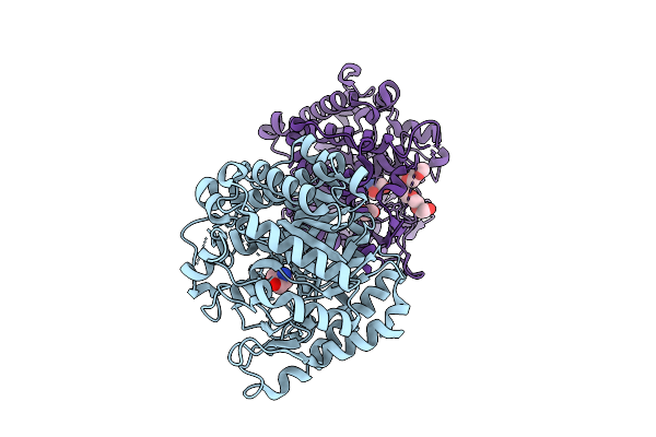 |
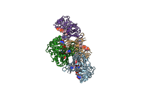
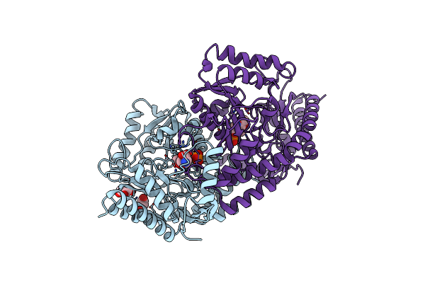
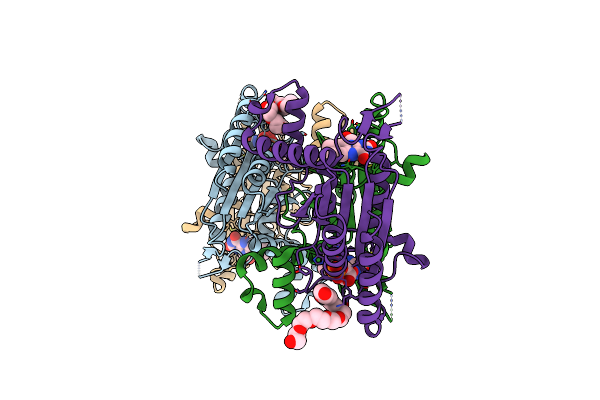
Crystal Structure Of Beta-Glucosidase From The Indigo-Producing Plant Polygonum Tinctorium
Organism: Persicaria tinctoria
Method: X-RAY DIFFRACTION Release Date: 2025-08-20 Classification: HYDROLASE Ligands: TRS, 1PG, EDO |
 |
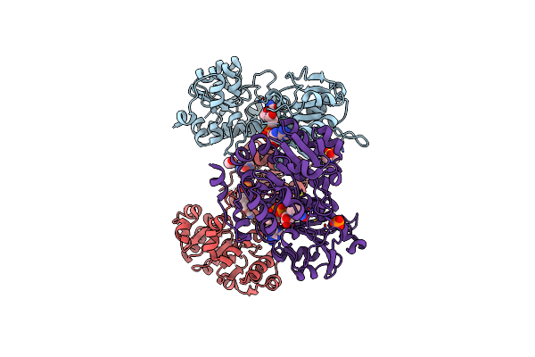
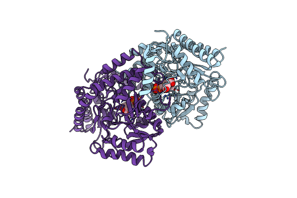
Crystal Structure Of Chitinase (E167Q) From The Carnivorous Plant Drosera Adelae
Organism: Drosera adelae
Method: X-RAY DIFFRACTION Release Date: 2025-08-13 Classification: HYDROLASE Ligands: ACY |
 |
Organism: Drosera adelae
Method: X-RAY DIFFRACTION Release Date: 2025-08-13 Classification: HYDROLASE Ligands: NAG, ACY |
 |
Organism: Saccharolobus solfataricus
Method: X-RAY DIFFRACTION Release Date: 2024-08-07 Classification: OXIDOREDUCTASE Ligands: NAD, 69O |
 |
Organism: Saccharolobus solfataricus
Method: X-RAY DIFFRACTION Release Date: 2024-08-07 Classification: OXIDOREDUCTASE Ligands: AKG, NAD, EDO |
 |
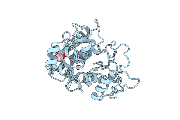
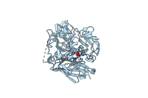
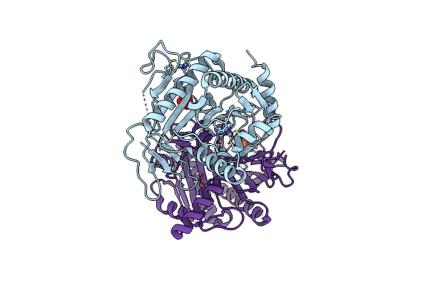
Crystal Structure Of Metal-Dependent Hydrolase Complexed With Manganese From Bacillus Smithii
Organism: Bacillus smithii
Method: X-RAY DIFFRACTION Resolution:2.53 Å Release Date: 2023-12-06 Classification: HYDROLASE Ligands: MN |
 |
Organism: Pseudomonas veronii
Method: X-RAY DIFFRACTION Resolution:2.20 Å Release Date: 2023-08-16 Classification: OXIDOREDUCTASE Ligands: NAP, LYS, IMD, EDO |
 |
Organism: Pseudomonas veronii
Method: X-RAY DIFFRACTION Resolution:2.50 Å Release Date: 2023-08-16 Classification: OXIDOREDUCTASE Ligands: NDP, ARG, EDO |
 |
Organism: Geobacillus kaustophilus
Method: X-RAY DIFFRACTION Resolution:2.20 Å Release Date: 2023-04-05 Classification: OXIDOREDUCTASE Ligands: NAD, EDO, EPE, PO4 |
 |
Organism: Geobacillus kaustophilus
Method: X-RAY DIFFRACTION Resolution:2.39 Å Release Date: 2023-04-05 Classification: OXIDOREDUCTASE Ligands: NAD, EDO, PYR |
 |
Crystal Structure Of Fmn-Dependent Nadph-Quinone Reductase (Azor) From Bacillus Cohnii
Organism: Bacillus cohnii
Method: X-RAY DIFFRACTION Resolution:1.57 Å Release Date: 2022-05-11 Classification: OXIDOREDUCTASE Ligands: FMN, IPA, GOL |
 |
Organism: Pyrococcus horikoshii (strain atcc 700860 / dsm 12428 / jcm 9974 / nbrc 100139 / ot-3)
Method: X-RAY DIFFRACTION Resolution:1.80 Å Release Date: 2022-03-30 Classification: TRANSFERASE Ligands: PLP, GOL, EDO |
 |
Organism: Pyrococcus horikoshii (strain atcc 700860 / dsm 12428 / jcm 9974 / nbrc 100139 / ot-3)
Method: X-RAY DIFFRACTION Resolution:1.92 Å Release Date: 2022-03-30 Classification: TRANSFERASE Ligands: PLP, ORN, GOL, EDO |
 |
Organism: Pyrococcus horikoshii (strain atcc 700860 / dsm 12428 / jcm 9974 / nbrc 100139 / ot-3)
Method: X-RAY DIFFRACTION Resolution:2.99 Å Release Date: 2022-03-30 Classification: TRANSFERASE Ligands: PGU |
 |
Organism: Pyrobaculum aerophilum (strain atcc 51768 / im2 / dsm 7523 / jcm 9630 / nbrc 100827)
Method: X-RAY DIFFRACTION Resolution:1.92 Å Release Date: 2020-05-20 Classification: OXIDOREDUCTASE Ligands: CU, C2O |
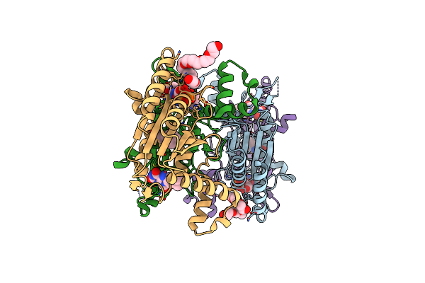 |
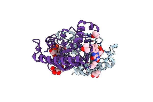
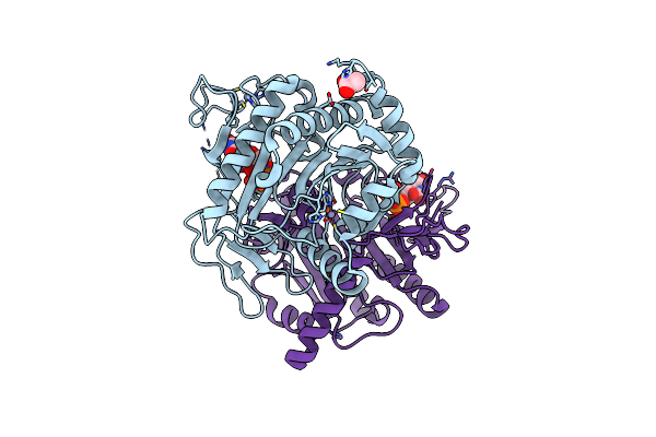
Crystal Structure Of Indigo Reductase From Bacillus Smithii Type Strain Dsm 4216
Organism: Bacillus smithii
Method: X-RAY DIFFRACTION Resolution:1.97 Å Release Date: 2020-04-01 Classification: OXIDOREDUCTASE Ligands: FMN, PE8, NHE |
 |
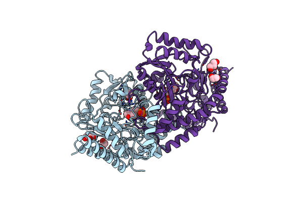
Crystal Structure Of Indigo Reductase (Y151F) From Bacillus Smithii Type Strain Dsm 4216
Organism: Bacillus smithii
Method: X-RAY DIFFRACTION Resolution:1.95 Å Release Date: 2020-04-01 Classification: OXIDOREDUCTASE Ligands: FMN, PE8 |
 |
Organism: Phytophthora infestans (strain t30-4)
Method: X-RAY DIFFRACTION Resolution:2.31 Å Release Date: 2020-04-01 Classification: OXIDOREDUCTASE Ligands: FLC, PE8, NAD |
 |
Organism: Pyrobaculum aerophilum str. im2
Method: X-RAY DIFFRACTION Resolution:2.33 Å Release Date: 2019-12-18 Classification: TRANSFERASE Ligands: ZN, FE, PO4 |
 |
Organism: Pyrobaculum aerophilum str. im2
Method: X-RAY DIFFRACTION Resolution:1.78 Å Release Date: 2019-12-18 Classification: TRANSFERASE Ligands: ZN, FE, ACT, UDP |