
Search Count: 48
 |
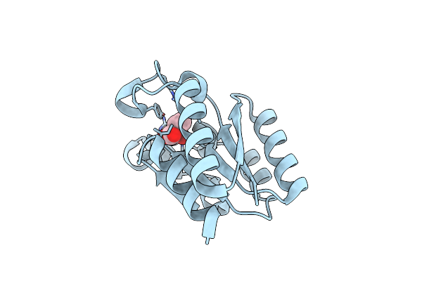
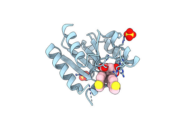
Organism: Homo sapiens
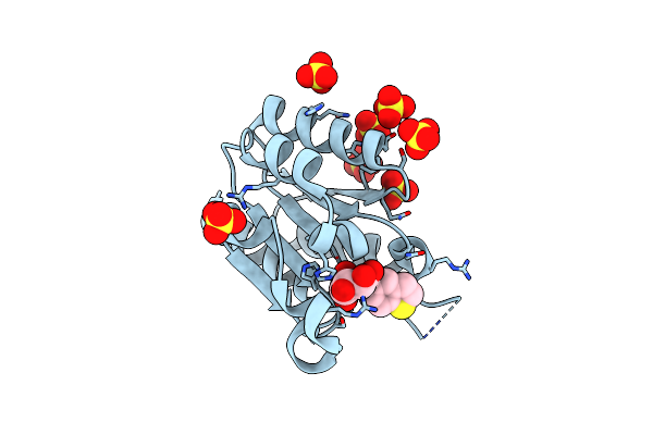
Method: X-RAY DIFFRACTION Resolution:1.78 Å Release Date: 2019-07-17 Classification: METAL BINDING PROTEIN Ligands: CA, P6G, NA, DMS, ACT, 1PE, FKW |
 |
Structure Of The Carboxy-Terminal Domain Of The Bacteriophage T5 L- Shaped Tail Fibre
Organism: Escherichia phage t5
Method: X-RAY DIFFRACTION Resolution:2.30 Å Release Date: 2015-12-16 Classification: VIRAL PROTEIN |
 |
Structure Of The Carboxy-Terminal Domain Of The Bacteriophage T5 L- Shaped Tail Fiber Without Its Intra-Molecular Chaperone Domain
Organism: Enterobacteria phage t5
Method: X-RAY DIFFRACTION Resolution:2.52 Å Release Date: 2015-08-05 Classification: VIRAL PROTEIN Ligands: GOL |
 |
Structure Of The Carboxy-Terminal Domain Of The Bacteriophage T5 L- Shaped Tail Fiber With Its Intra-Molecular Chaperone Domain
Organism: Enterobacteria phage t5
Method: X-RAY DIFFRACTION Resolution:2.52 Å Release Date: 2015-08-05 Classification: VIRAL PROTEIN Ligands: FLC |
 |
Structure Of The Mycobacterium Tuberculosis Type Ii Dehydroquinase N12S Mutant (Crystal Form 1)
Organism: Mycobacterium tuberculosis
Method: X-RAY DIFFRACTION Resolution:2.70 Å Release Date: 2015-03-25 Classification: LYASE Ligands: GOL |
 |
Structure Of The Mycobacterium Tuberculosis Type Ii Dehydroquinase N12S Mutant (Crystal Form 2)
Organism: Mycobacterium tuberculosis
Method: X-RAY DIFFRACTION Resolution:2.60 Å Release Date: 2015-03-25 Classification: LYASE Ligands: TRS |
 |
Structure Of The Mycobacterium Tuberculosis Type Ii Dehydroquinase Inhibited By A 3-Dehydroquinic Acid Derivative
Organism: Mycobacterium tuberculosis
Method: X-RAY DIFFRACTION Resolution:1.65 Å Release Date: 2015-03-25 Classification: LYASE |
 |
Structure Of The Mycobacterium Tuberculosis Type Ii Dehydroquinase D88N Mutant
Organism: Mycobacterium tuberculosis
Method: X-RAY DIFFRACTION Resolution:2.52 Å Release Date: 2015-03-25 Classification: LYASE |
 |
Structure Of The Mycobacterium Tuberculosis Type Ii Dehydroquinase Inhibited By A 3-Dehydroquinic Acid Derivative
Organism: Mycobacterium tuberculosis
Method: X-RAY DIFFRACTION Resolution:3.10 Å Release Date: 2015-03-25 Classification: LYASE Ligands: 9PY |
 |
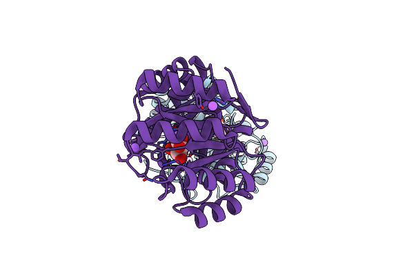
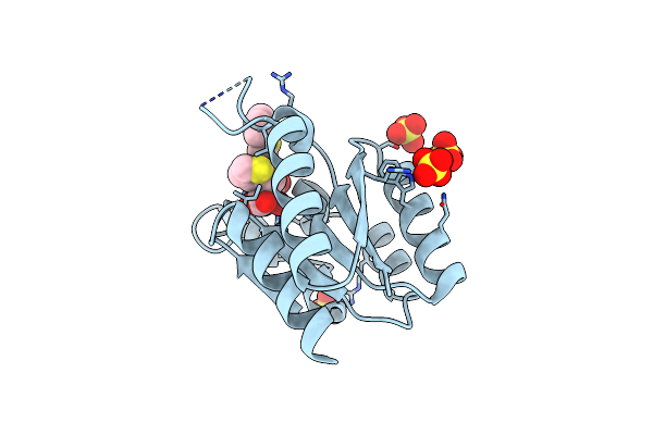
Organism: Salmonella enterica subsp. enterica serovar typhi
Method: X-RAY DIFFRACTION Resolution:1.00 Å Release Date: 2015-02-18 Classification: LYASE |
 |
Structure Of The Salmonella Typhi Type I Dehydroquinase Inhibited By A 3-Epiquinic Acid Derivative
Organism: Salmonella enterica subsp. enterica serovar typhi
Method: X-RAY DIFFRACTION Resolution:1.15 Å Release Date: 2015-02-18 Classification: LYASE Ligands: 9C4, NA, CL |
 |
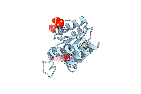
Crystal Structure Of Mycobacterium Tuberculosis Shikimate Kinase In Complex With Adp And A Shikimic Acid Derivative.
Organism: Mycobacterium tuberculosis
Method: X-RAY DIFFRACTION Resolution:2.15 Å Release Date: 2013-08-07 Classification: TRANSFERASE Ligands: ADP, K2Q |
 |
Structure Of Mycobacterium Tuberculosis Type Ii Dehydroquinase Inhibited By (2S)-2-(4-Methoxy)Benzyl-3-Dehydroquinic Acid
Organism: Mycobacterium tuberculosis
Method: X-RAY DIFFRACTION Resolution:2.00 Å Release Date: 2012-12-19 Classification: LYASE Ligands: 3DQ, SO4 |
 |
Structure Of Mycobacterium Tuberculosis Type Ii Dehydroquinase Inhibited By (2S)-2-Perfluorobenzyl-3-Dehydroquinic Acid
Organism: Mycobacterium tuberculosis
Method: X-RAY DIFFRACTION Resolution:2.30 Å Release Date: 2012-12-19 Classification: LYASE Ligands: 2HN, SO4 |
 |
Structure Of Mycobacterium Tuberculosis Type Ii Dehydroquinase Inhibited By (2R)-2-(Benzothiophen-5-Yl)Methyl-3-Dehydroquinic Acid
Organism: Mycobacterium tuberculosis
Method: X-RAY DIFFRACTION Resolution:1.54 Å Release Date: 2012-12-19 Classification: LYASE Ligands: BZ5, SO4 |
 |
Structure Of Helicobacter Pylori Type Ii Dehydroquinase Inhibited By (2S)-2-(4-Methoxy)Benzyl-3-Dehydroquinic Acid
Organism: Helicobacter pylori 26695
Method: X-RAY DIFFRACTION Resolution:2.00 Å Release Date: 2012-12-19 Classification: LYASE Ligands: 3DQ, SO4 |
 |
Structure Of Helicobacter Pylori Type Ii Dehydroquinase Inhibited By (2S)-2-Perfluorobenzyl-3-Dehydroquinic Acid
Organism: Helicobacter pylori 26695
Method: X-RAY DIFFRACTION Resolution:1.90 Å Release Date: 2012-12-19 Classification: LYASE Ligands: 2HN, PO4 |
 |
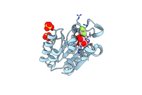
Structure Of Mycobacterium Tuberculosis Type Ii Dehydroquinase Complexed With (1R,4S,5R)-1,4,5-Trihydroxy-3-((5-Methylbenzo(B) Thiophen-2-Yl)Methoxy)Cyclohex-2-Enecarboxylate
Organism: Mycobacterium tuberculosis
Method: X-RAY DIFFRACTION Resolution:1.50 Å Release Date: 2011-08-17 Classification: LYASE Ligands: CB6, NA, SO4 |
 |
Structure Of Mycobacterium Tuberculosis Type Ii Dehydroquinase Complexed With (1R,4S,5R)-3-(Benzo(B)Thiophen-5-Ylmethoxy)-2-(Benzo(B) Thiophen-5-Ylmethyl)-1,4,5-Trihydroxycyclohex-2-Enecarboxylate
Organism: Mycobacterium tuberculosis
Method: X-RAY DIFFRACTION Resolution:2.50 Å Release Date: 2011-08-17 Classification: LYASE Ligands: CB7, SO4 |
 |
Structure Of Mycobacterium Tuberculosis Type Ii Dehydroquinase Complexed With (1R,4S,5R)-3-(Benzo(B)Thiophen-2-Ylmethoxy)-1,4,5- Trihydroxy-2-(Thiophen-2-Ylmethyl)Cyclohex-2-Enecarboxylate
Organism: Mycobacterium tuberculosis
Method: X-RAY DIFFRACTION Resolution:1.50 Å Release Date: 2011-08-17 Classification: LYASE Ligands: CB8, SO4 |