
Search Count: 28
 |
Organism: Mus musculus
Method: X-RAY DIFFRACTION Resolution:1.80 Å Release Date: 2020-08-26 Classification: UNKNOWN FUNCTION Ligands: ZN |
 |
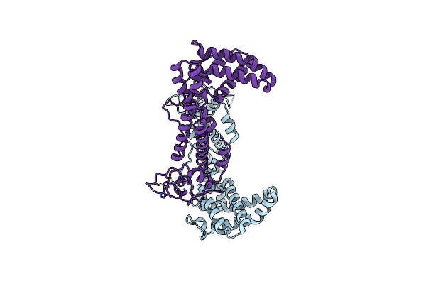
The X-Ray Structure Of Yeast Trna Methyltransferase Complex Of Trm7 And Trm734 Essential For 2'-O-Methylation At The First Position Of Anticodon In Specific Trnas
Organism: Saccharomyces cerevisiae s288c
Method: X-RAY DIFFRACTION Resolution:2.70 Å Release Date: 2019-10-02 Classification: TRANSFERASE Ligands: SO4, EPE |
 |
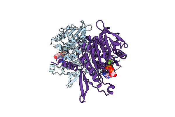
The X-Ray Structure Of Yeast Trna Methyltransferase Trm7-Trm734 In Complex With S-Adenosyl-L-Methionine
Organism: Saccharomyces cerevisiae s288c
Method: X-RAY DIFFRACTION Resolution:2.32 Å Release Date: 2019-10-02 Classification: TRANSFERASE Ligands: EPE, SO4, SAM |
 |
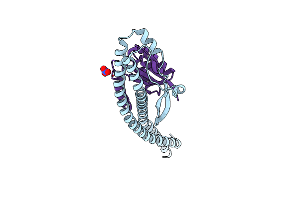
The Crystal Structure Of The Minimal Core Domain Of The Microtubule Depolymerizer Kif2C Complexed With Adp-Mg-Alfx
Organism: Mus musculus
Method: X-RAY DIFFRACTION Resolution:3.43 Å Release Date: 2017-09-13 Classification: STRUCTURAL PROTEIN Ligands: MG, ADP, AF3 |
 |
The Crystal Structure Of The Minimal Core Domain Of The Microtubule Depolymerizer Kif2C Complexed With Adp-Mg-Befx
Organism: Mus musculus
Method: X-RAY DIFFRACTION Resolution:3.10 Å Release Date: 2017-09-13 Classification: STRUCTURAL PROTEIN Ligands: ADP, BEF, MG |
 |
Organism: Homo sapiens
Method: X-RAY DIFFRACTION Resolution:2.10 Å Release Date: 2017-06-28 Classification: ONCOPROTEIN |
 |
Organism: Homo sapiens
Method: X-RAY DIFFRACTION Resolution:3.20 Å Release Date: 2016-07-27 Classification: HYDROLASE Ligands: CA |
 |
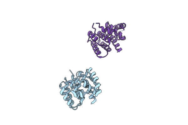
Crystal Structure Analysis Of The Mini-Chaperonines, Variant With Gly 184 Replaced With Ile And Leu 185 Replaced Val And Val 186 Replaced With Leu.
Organism: Escherichia coli
Method: X-RAY DIFFRACTION Resolution:1.50 Å Release Date: 2013-11-13 Classification: CHAPERONE |
 |
Organism: Escherichia coli
Method: X-RAY DIFFRACTION Resolution:1.80 Å Release Date: 2013-11-13 Classification: CHAPERONE |
 |
Crystal Structure Analysis Of The Mini-Chaperonin Variant With Leu 185, Val 186, Pro 187, Arg 188 And Ser 190 Replaced With All Gly
Organism: Escherichia coli
Method: X-RAY DIFFRACTION Resolution:1.90 Å Release Date: 2013-11-13 Classification: CHAPERONE |
 |
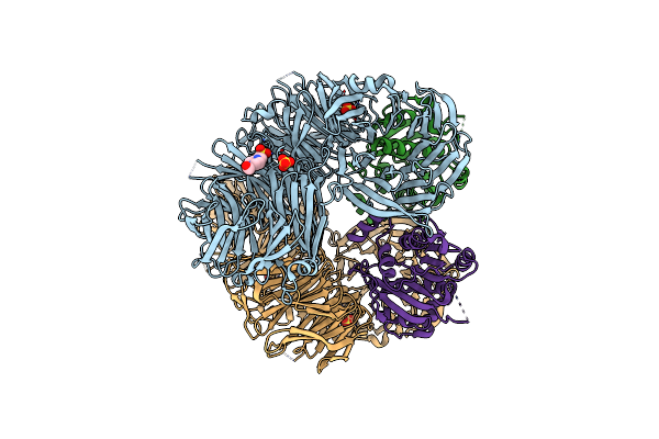
Crystal Structure Of Nucleotide-Free A3B3 Complex From Enterococcus Hirae V-Atpase [Ea3B3]
Organism: Enterococcus hirae
Method: X-RAY DIFFRACTION Resolution:2.80 Å Release Date: 2013-01-16 Classification: HYDROLASE |
 |
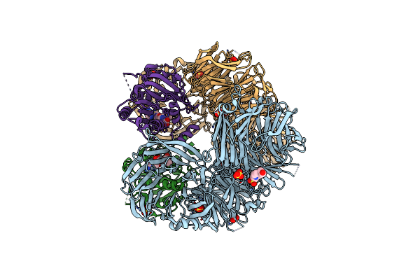
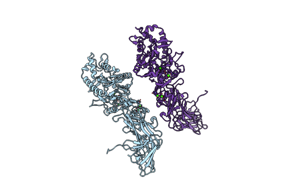
Crystal Structure Of Amp-Pnp Bound A3B3 Complex From Enterococcus Hirae V-Atpase [Ba3B3]
Organism: Enterococcus hirae
Method: X-RAY DIFFRACTION Resolution:3.40 Å Release Date: 2013-01-16 Classification: HYDROLASE Ligands: ANP, MG |
 |
Organism: Enterococcus hirae
Method: X-RAY DIFFRACTION Resolution:2.17 Å Release Date: 2013-01-16 Classification: HYDROLASE Ligands: GOL, CL, B3P |
 |
Organism: Enterococcus hirae
Method: X-RAY DIFFRACTION Resolution:3.90 Å Release Date: 2013-01-16 Classification: HYDROLASE |
 |
Organism: Enterococcus hirae
Method: X-RAY DIFFRACTION Resolution:2.68 Å Release Date: 2013-01-16 Classification: HYDROLASE Ligands: ANP, MG |
 |
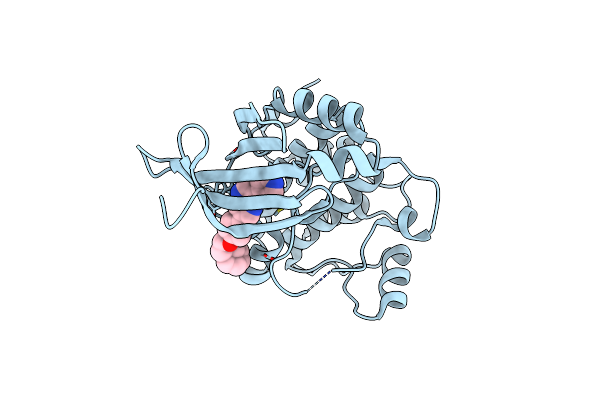
Crystal Structure Of Aeromonas Proteolytica Aminopeptidase Complexed With 8-Quinolinol
Organism: Vibrio proteolyticus
Method: X-RAY DIFFRACTION Resolution:1.29 Å Release Date: 2012-05-02 Classification: HYDROLASE/HYDROLASE INHIBITOR Ligands: ZN, HQY, NA, CL, SCN, GOL |
 |
Crystal Structure Of The Central Axis (Ntpd-Ntpg) In The Catalytic Portion Of Enterococcus Hirae V-Type Sodium Atpase
Organism: Enterococcus hirae
Method: X-RAY DIFFRACTION Resolution:2.00 Å Release Date: 2011-10-05 Classification: HYDROLASE Ligands: NO3 |
 |
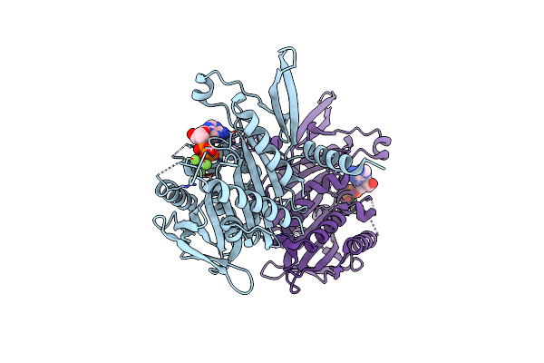
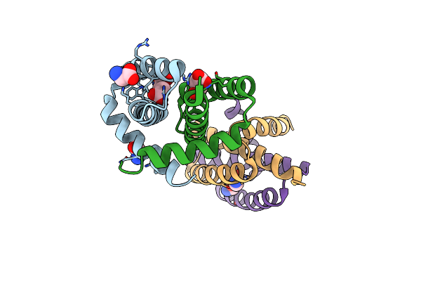
Human Amp-Activated Protein Kinase Alpha 2 Subunit Kinase Domain (T172D) Complexed With Compound C
Organism: Homo sapiens
Method: X-RAY DIFFRACTION Resolution:2.08 Å Release Date: 2011-04-27 Classification: TRANSFERASE/TRANSFERASE INHIBITOR Ligands: TAK |
 |
Crystal Structure Of Rna-Binding Domain Of Ns1 From Influenza A Virus A/Crow/Kyoto/T1/2004(H5N1)
Organism: Influenza a virus
Method: X-RAY DIFFRACTION Resolution:1.85 Å Release Date: 2008-05-13 Classification: VIRAL PROTEIN Ligands: GLY, SIN |
 |
Crystal Structure Of Matrix Protein 1 From Influenza A Virus A/Crow/Kyoto/T1/2004(H5N1)
Organism: Influenza a virus
Method: X-RAY DIFFRACTION Resolution:2.02 Å Release Date: 2008-05-13 Classification: VIRAL PROTEIN |