
Search Count: 17
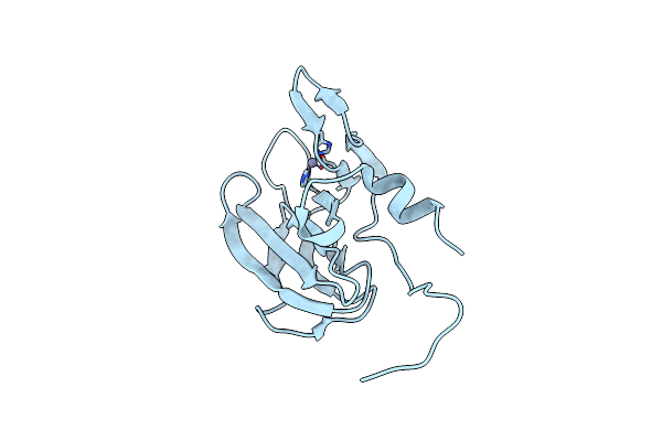 |
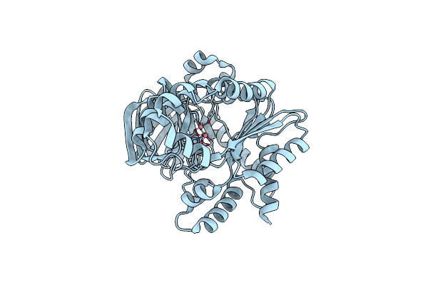
Organism: Streptococcus thermophilus
Method: X-RAY DIFFRACTION Resolution:1.35 Å Release Date: 2025-06-04 Classification: HYDROLASE Ligands: ZN |
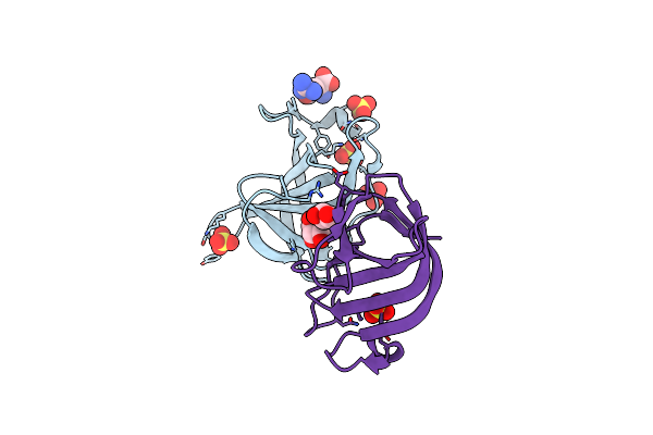 |
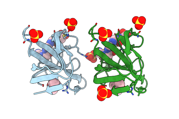
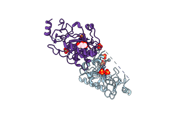
Organism: Staphylococcus simulans
Method: X-RAY DIFFRACTION Resolution:1.47 Å Release Date: 2021-02-17 Classification: CELL ADHESION Ligands: GOL, GGB, SO4 |
 |
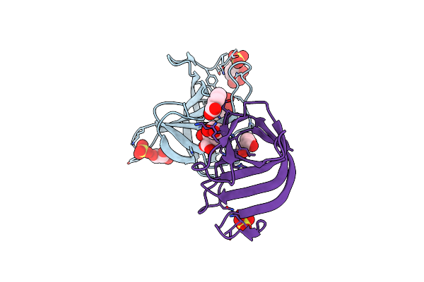
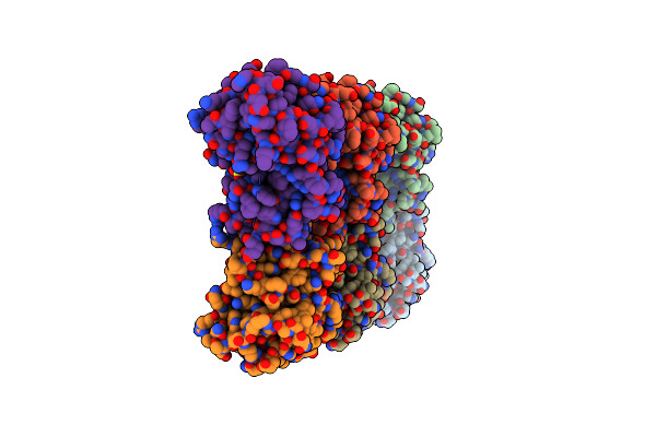
Organism: Staphylococcus simulans
Method: X-RAY DIFFRACTION Resolution:0.84 Å Release Date: 2021-02-17 Classification: CELL ADHESION Ligands: BEZ, GOL, SO4, NA, MES |
 |
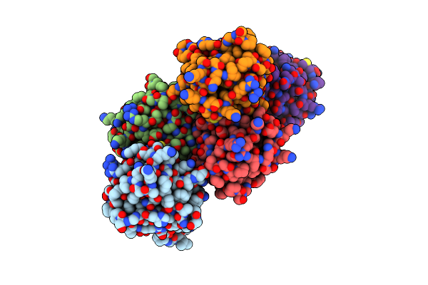
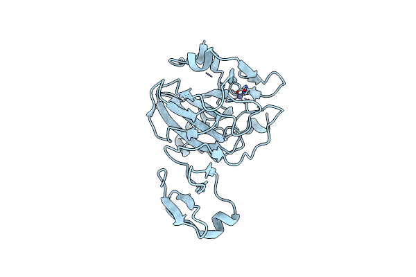
Organism: Staphylococcus simulans
Method: X-RAY DIFFRACTION Resolution:1.31 Å Release Date: 2021-02-17 Classification: CELL ADHESION Ligands: SAL, GOL, SO4 |
 |
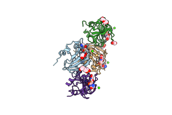
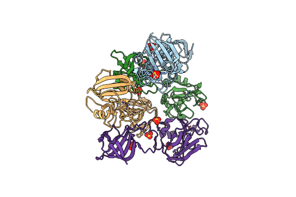
Crystal Structure Of Catalytic Domain A109H Mutant Of Prophage-Encoded M23 Protein Enpa From Enterococcus Faecalis.
Organism: Enterococcus faecalis (strain atcc 700802 / v583)
Method: X-RAY DIFFRACTION Resolution:3.00 Å Release Date: 2020-09-09 Classification: ANTIMICROBIAL PROTEIN Ligands: ZN |
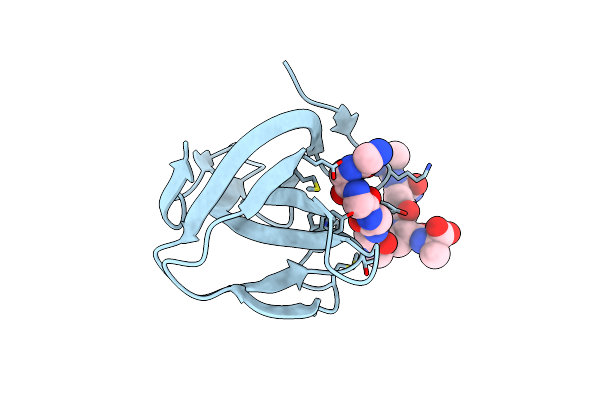 |
Organism: Staphylococcus simulans
Method: X-RAY DIFFRACTION Resolution:2.50 Å Release Date: 2019-10-16 Classification: PEPTIDE BINDING PROTEIN Ligands: K5T |
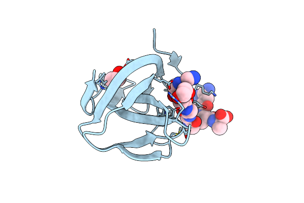 |
Organism: Staphylococcus simulans
Method: X-RAY DIFFRACTION Resolution:1.43 Å Release Date: 2019-10-16 Classification: PEPTIDE BINDING PROTEIN Ligands: K5T, EDO |
 |
Organism: Staphylococcaceae
Method: X-RAY DIFFRACTION Resolution:1.60 Å Release Date: 2017-07-12 Classification: CELL ADHESION Ligands: SO4, MPD |
 |
Organism: Staphylococcus aureus subsp. aureus nctc 8325
Method: X-RAY DIFFRACTION Resolution:1.50 Å Release Date: 2015-10-21 Classification: HYDROLASE Ligands: ZN, CA, CL, UNL, EDO, 4SQ, EPE, PEG |
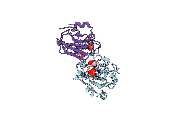 |
Catalytic Domain Of The Antimicrobial Peptidase Lysostaphin From Staphylococcus Simulans Crystallized In The Presence Of Phosphate
Organism: Staphylococcus simulans bv. staphylolyticus
Method: X-RAY DIFFRACTION Resolution:1.26 Å Release Date: 2014-07-16 Classification: HYDROLASE Ligands: ZN, PO4, GOL |
 |
Organism: Staphylococcus simulans
Method: X-RAY DIFFRACTION Resolution:3.50 Å Release Date: 2014-07-09 Classification: HYDROLASE Ligands: ZN, SO4 |
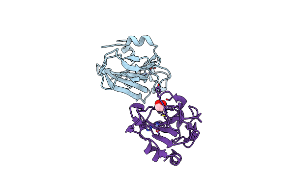 |
Catalytic Domain Of The Antimicrobial Peptidase Lysostaphin From Staphylococcus Simulans Crystallized In The Absence Of Phosphate
Organism: Staphylococcus simulans
Method: X-RAY DIFFRACTION Resolution:1.78 Å Release Date: 2014-07-09 Classification: HYDROLASE Ligands: ZN, EDO |
 |
Organism: Thermus thermophilus
Method: X-RAY DIFFRACTION Resolution:3.70 Å Release Date: 2005-11-08 Classification: HYDROLASE Ligands: ZN |
 |
Organism: Staphylococcus aureus subsp. aureus
Method: X-RAY DIFFRACTION Resolution:1.80 Å Release Date: 2005-06-07 Classification: HYDROLASE Ligands: CO |
 |
Organism: Escherichia coli
Method: X-RAY DIFFRACTION Resolution:1.40 Å Release Date: 2004-09-07 Classification: HYDROLASE Ligands: SO4, BU1 |
 |
Organism: Escherichia coli
Method: X-RAY DIFFRACTION Resolution:2.40 Å Release Date: 2004-09-07 Classification: HYDROLASE Ligands: ZN, SO4 |
 |
Organism: Staphylococcus aureus subsp. aureus nctc 8325
Method: X-RAY DIFFRACTION Resolution:1.30 Å Release Date: 2004-01-20 Classification: HYDROLASE Ligands: ZN |