
Search Count: 16
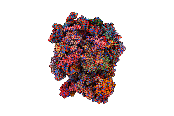 |
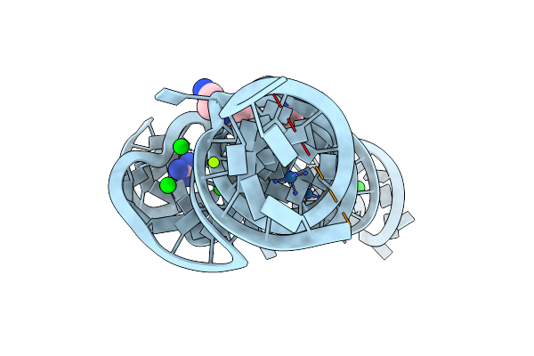
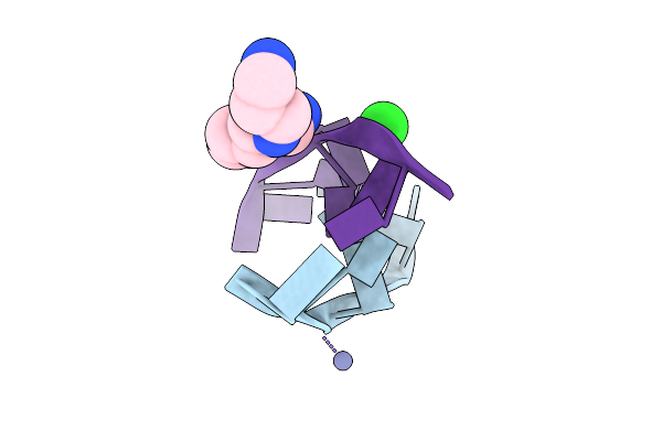
Structure Of Candida Albicans 80S Ribosome In Complex With Mefloquine (Non-Rotated State)
Organism: Candida albicans sc5314
Method: ELECTRON MICROSCOPY Release Date: 2025-04-23 Classification: RIBOSOME Ligands: ZN, YMZ, SPK |
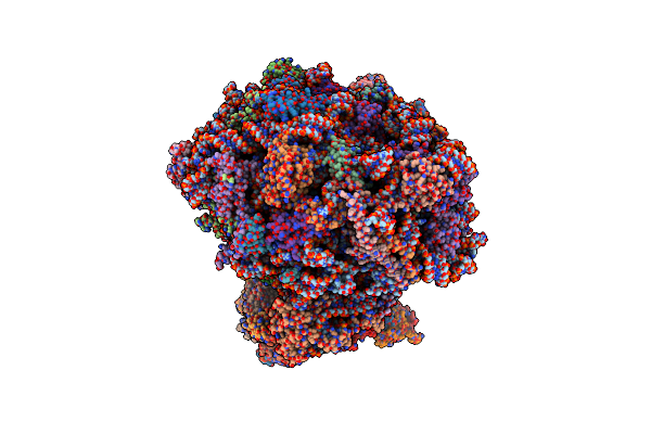 |
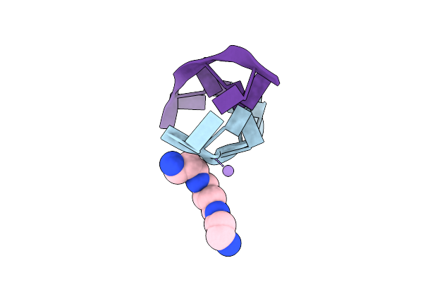
The Structure Of The Candida Albicans Ribosome With Trna-Fmet, Mrna, And Compounds (Gen And Mfq) Shows Strong Density For The A Site Trna
Organism: Candida albicans
Method: ELECTRON MICROSCOPY Release Date: 2025-04-23 Classification: RIBOSOME Ligands: SPK, GET, ZN |
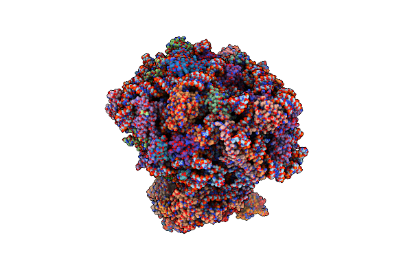 |
The Structure Of The Candida Albicans Ribosome With Trna-Fmet, Mrna, And Compounds (Gen And Mfq) With Strong Density For The P-Site Trna
Organism: Escherichia coli, Candida albicans sc5314
Method: ELECTRON MICROSCOPY Release Date: 2025-04-23 Classification: RIBOSOME Ligands: SPK, GET, ZN, YMZ |
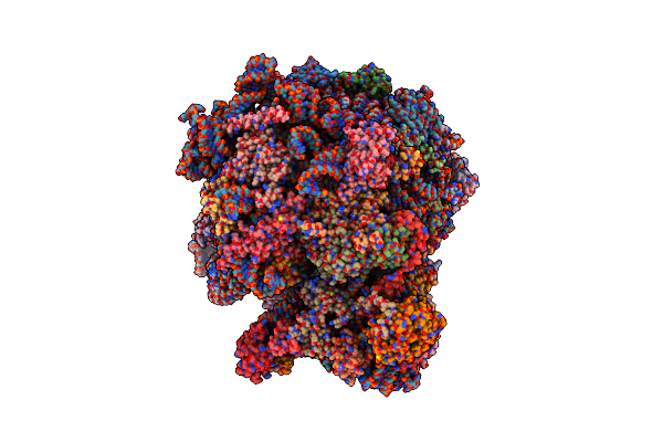 |
Organism: Candida albicans
Method: ELECTRON MICROSCOPY Release Date: 2024-10-02 Classification: RIBOSOME Ligands: YMZ, SPK, MG, ZN |
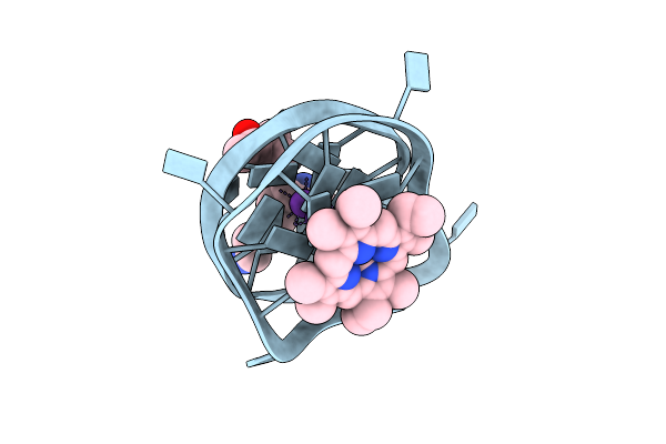 |
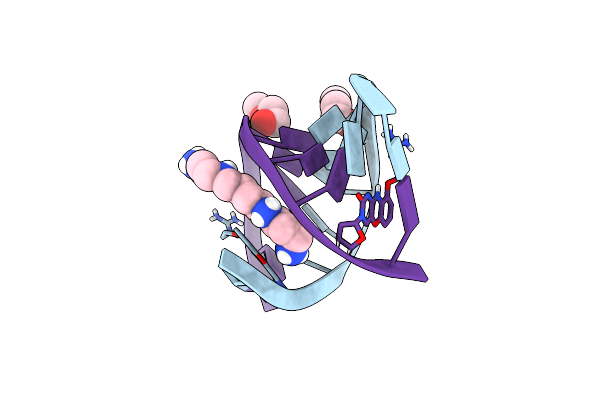
Right-Left Hybrid Parallel G-Quadruplex In Complex With N-Methyl Mesoporphyrin
Organism: Homo sapiens
Method: X-RAY DIFFRACTION Resolution:1.45 Å Release Date: 2024-07-03 Classification: DNA Ligands: SPK, MPD, MMP, K |
 |
Organism: Synthetic construct, Candida albicans
Method: ELECTRON MICROSCOPY Release Date: 2023-09-13 Classification: RIBOSOME Ligands: SPK, K16, SPD, MG, ZN |
 |
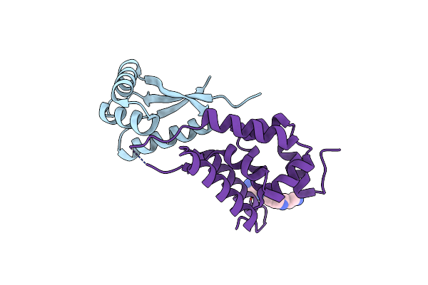
Human Atp13A2 In The Outward-Facing E2 State Bound To Spermine And Beryllium Fluoride
Organism: Homo sapiens
Method: ELECTRON MICROSCOPY Release Date: 2022-03-30 Classification: TRANSPORT PROTEIN Ligands: MG, BEF, Y01, LMT, CLR, D10, C14, D12, NAG, SPK, EUJ |
 |
Human Atp13A2 In The Outward-Facing E2 State Bound To Spermine And Magnesium Fluoride
Organism: Homo sapiens
Method: ELECTRON MICROSCOPY Release Date: 2022-03-30 Classification: TRANSPORT PROTEIN Ligands: MG, MF4, Y01, LMT, CLR, D10, C14, D12, NAG, SPK, EUJ |
 |
Organism: Sulfobacillus acidophilus dsm 10332
Method: X-RAY DIFFRACTION Resolution:2.71 Å Release Date: 2017-01-11 Classification: RNA Ligands: SR, SPK, MG, IRI, GAI |
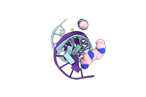 |
The Atomic Structure Of 5-Hydroxymethyl 2'-Deoxycitidine Base Paired With 2'-Deoxyguanosine In Dickerson Drew Dodecamer
Organism: Synthetic dna
Method: X-RAY DIFFRACTION Resolution:1.02 Å Release Date: 2013-11-20 Classification: DNA Ligands: MG, SPK |
 |
Organism: Synthetic construct
Method: X-RAY DIFFRACTION Resolution:0.85 Å Release Date: 2013-06-05 Classification: DNA Ligands: ZN, CL, SPK |
 |
Organism: Synthetic construct
Method: X-RAY DIFFRACTION Resolution:0.75 Å Release Date: 2013-06-05 Classification: DNA Ligands: SPK, MN |
 |
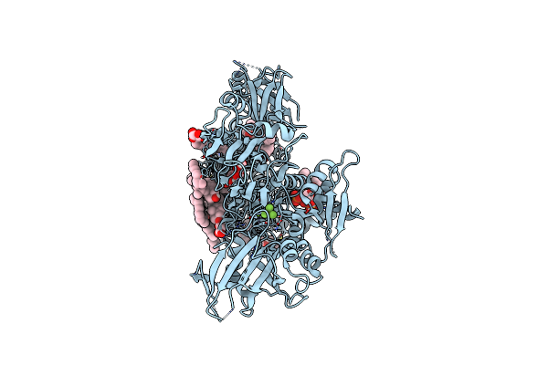
Crystal Structure Of Streptomyces Coelicolor A3(2) Cyp158A2 From Antibiotic Biosynthetic Pathways
Organism: Streptomyces coelicolor
Method: X-RAY DIFFRACTION Resolution:1.75 Å Release Date: 2005-01-25 Classification: OXIDOREDUCTASE Ligands: SPK, HEM, MES |
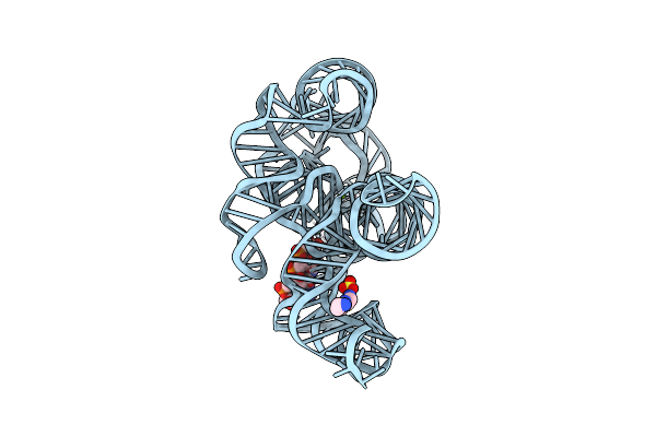 |
Organism: Staphylococcus phage twort
Method: X-RAY DIFFRACTION Resolution:3.60 Å Release Date: 2004-12-21 Classification: RNA Ligands: SPK, MG, SO4 |
 |
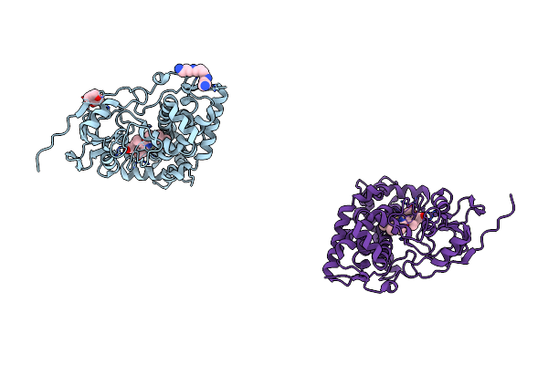
Organism: Escherichia coli
Method: X-RAY DIFFRACTION Resolution:2.30 Å Release Date: 2002-11-27 Classification: CHAPERONE Ligands: SPK |
 |
Direct Observation Of A Cytosine Analog That Forms Five Hydrogen Bonds To Guanosine; Guanyl G-Clamp
Method: X-RAY DIFFRACTION
Resolution:1.00 Å Release Date: 2001-12-21 Classification: DNA Ligands: MOE, SPK |