
Search Count: 24
 |
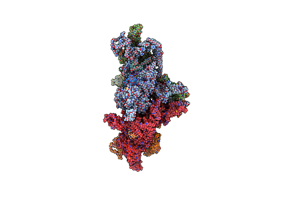
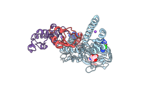
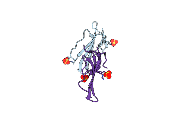
Co-Crystal Of Broadly Neutralizing Biparatopic Vhh In Complex With Cardiotoxin (P01468) Naja Pallida
Organism: Vicugna pacos, Naja pallida
Method: X-RAY DIFFRACTION Release Date: 2025-11-05 Classification: TOXIN Ligands: ACT |
 |
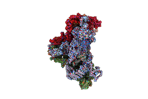
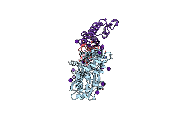
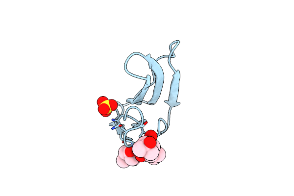
Co-Crystal Of Broadly Neutralizing Vhh In Complex With Short Neurotoxin 1 (P01426) Naja Pallida
Organism: Vicugna pacos, Naja pallida
Method: X-RAY DIFFRACTION Release Date: 2025-11-05 Classification: TOXIN Ligands: NI |
 |
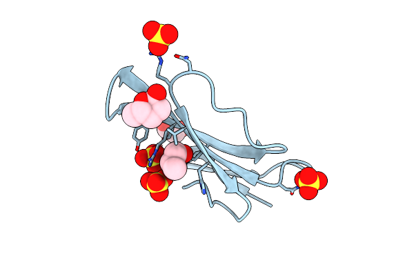
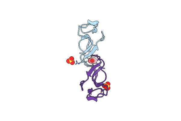
Organism: Vicugna pacos, Naja kaouthia
Method: X-RAY DIFFRACTION Release Date: 2025-08-06 Classification: TOXIN Ligands: HOH |
 |
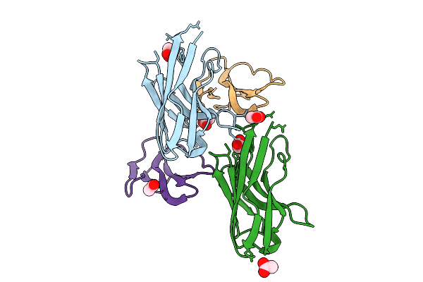
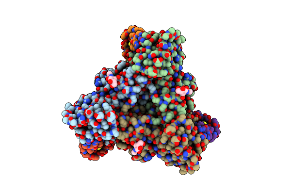
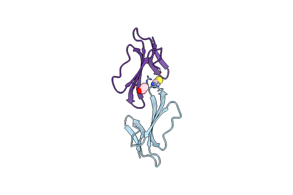
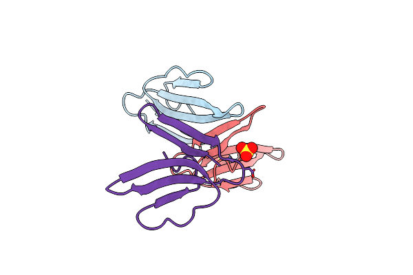
Crystal Structure Of Long Neurotoxin From The Venom Of The King Cobra (3Ftx-L15) In Complex With Fab Of Broadly Neutralizing Antibody 95Mat5
Organism: Homo sapiens, Ophiophagus hannah
Method: X-RAY DIFFRACTION Resolution:2.90 Å Release Date: 2024-03-20 Classification: TOXIN/IMMUNE SYSTEM |
 |
Organism: Oryctolagus cuniculus
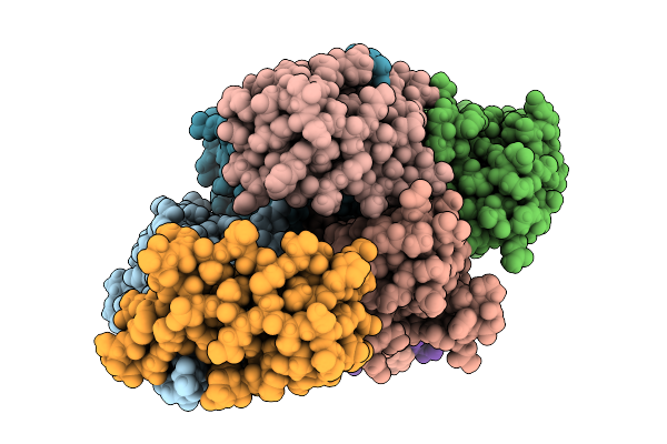
Method: ELECTRON MICROSCOPY Release Date: 2023-05-31 Classification: MEMBRANE PROTEIN Ligands: CFF, ATP, ZN |
 |
Focus/Local Refined Map In C1 Of Signal Subtracted Ryr1 Particles In Complex With Imperacalcin
Organism: Pandinus imperator, Oryctolagus cuniculus
Method: ELECTRON MICROSCOPY Resolution:3.14 Å Release Date: 2023-05-31 Classification: TOXIN Ligands: ATP, CFF |
 |
Organism: Homo sapiens, Pandinus imperator, Oryctolagus cuniculus
Method: ELECTRON MICROSCOPY Release Date: 2023-05-31 Classification: MEMBRANE PROTEIN Ligands: CFF, CA, ATP, ZN |
 |
Organism: Homo sapiens, Oryctolagus cuniculus
Method: ELECTRON MICROSCOPY Release Date: 2023-05-31 Classification: MEMBRANE PROTEIN Ligands: CFF, CA, ATP, ZN |
 |
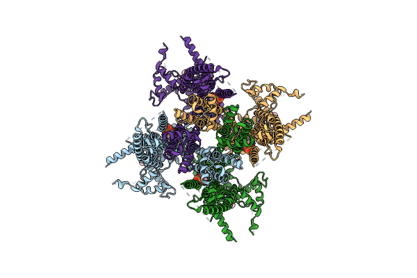
Cryo-Em Strucutre Of Human Acid-Sensing Ion Channel 1A In Complex With Snake Toxin Mambalgin1 At Ph 8.0
Organism: Homo sapiens, Dendroaspis polylepis polylepis
Method: ELECTRON MICROSCOPY Release Date: 2020-10-21 Classification: MEMBRANE PROTEIN Ligands: NAG |
 |
Organism: Dendroaspis angusticeps
Method: X-RAY DIFFRACTION Resolution:1.80 Å Release Date: 2017-05-03 Classification: TOXIN Ligands: PGO, EDO, SO4, YVR |
 |
Organism: Gallus gallus, Micrurus tener tener
Method: X-RAY DIFFRACTION Resolution:2.07 Å Release Date: 2014-02-19 Classification: TRANSPORT PROTEIN/TOXIN Ligands: NAG, CL, NA, P6G |
 |
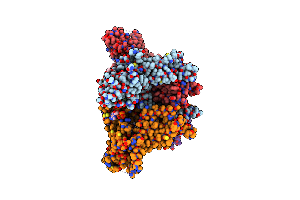
Structure Of Acid-Sensing Ion Channel In Complex With Snake Toxin And Amiloride
Organism: Gallus gallus, Micrurus tener tener
Method: X-RAY DIFFRACTION Resolution:2.27 Å Release Date: 2014-02-19 Classification: TRANSPORT PROTEIN/TOXIN Ligands: NAG, CL, NA, P6G, AMR |
 |
Cesium Sites In The Crystal Structure Of Acid-Sensing Ion Channel In Complex With Snake Toxin
Organism: Gallus gallus, Micrurus tener tener
Method: X-RAY DIFFRACTION Resolution:2.65 Å Release Date: 2014-02-19 Classification: TRANSPORT PROTEIN/TOXIN Ligands: CS, CL, PE4 |
 |
Organism: Dendroaspis angusticeps
Method: X-RAY DIFFRACTION Resolution:1.80 Å Release Date: 2012-06-27 Classification: TOXIN Ligands: SCN, ACT |
 |
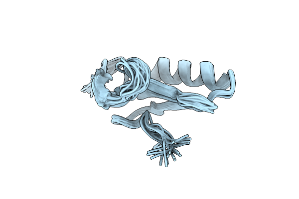
Expression In Pichia Pastoris And Backbone Dynamics Of Dendroaspin, A Three Finger Toxin
Organism: Dendroaspis jamesoni kaimosae
Method: SOLUTION NMR Release Date: 2012-03-07 Classification: TOXIN |
 |
Crystal Structure Of The Chimeric Muscarinic Toxin Mt7 With Loop 3 From Mt1
Organism: Dendroaspis angusticeps
Method: X-RAY DIFFRACTION Resolution:1.25 Å Release Date: 2011-08-24 Classification: TOXIN Ligands: SO4 |
 |
Method: X-RAY DIFFRACTION
Resolution:1.20 Å Release Date: 2010-08-11 Classification: TOXIN Ligands: MPD, SO4 |
 |
Crystal Structure Of The Chimeric Muscarinic Toxin Mt7 With Loop 1 From Mt1.
Organism: Dendroaspis angusticeps
Method: X-RAY DIFFRACTION Resolution:1.30 Å Release Date: 2009-12-22 Classification: TOXIN Ligands: SO4 |
 |
Organism: Dendroaspis angusticeps
Method: X-RAY DIFFRACTION Resolution:1.39 Å Release Date: 2008-10-14 Classification: TOXIN Ligands: ACT, SO4 |
 |
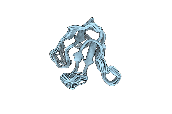
Solution Structure Of The Extracellular Domain Of Prod1, A Protein Implicated In Proximodistal Identity During Amphibian Limb Regeneration
Organism: Notophthalmus viridescens
Method: SOLUTION NMR Release Date: 2008-09-30 Classification: TOXIN |