
Search Count: 1,048
All
Selected
 |
Organism: Homo sapiens
Method: ELECTRON MICROSCOPY Resolution:2.50 Å Release Date: 2026-01-28 Classification: LIGASE Ligands: ZN, A1CED |
 |
Organism: Homo sapiens
Method: X-RAY DIFFRACTION Resolution:2.80 Å Release Date: 2026-01-14 Classification: SIGNALING PROTEIN |
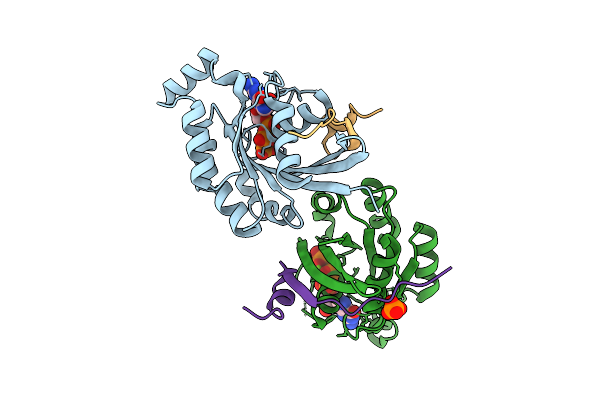 |
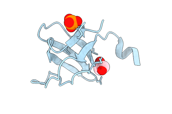
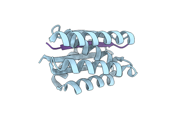
Crystal Structure Of Human Rac1 In Complex With The Scaffold Protein Posh (Residues 321-348)
Organism: Homo sapiens
Method: X-RAY DIFFRACTION Resolution:1.85 Å Release Date: 2025-12-03 Classification: HYDROLASE Ligands: GNP, MG, PO4, CL |
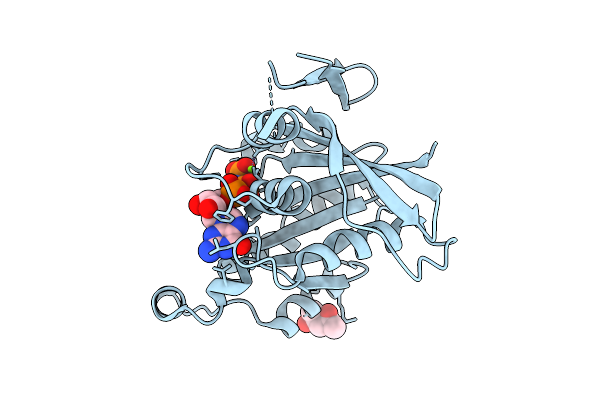 |
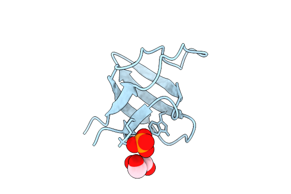
Crystal Structure Of Human Rac1 Fused With The Scaffold Protein Posh (Residues 319-371)
Organism: Homo sapiens
Method: X-RAY DIFFRACTION Resolution:1.25 Å Release Date: 2025-12-03 Classification: HYDROLASE Ligands: GNP, MG, MPD |
 |
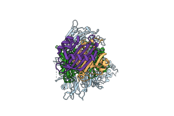
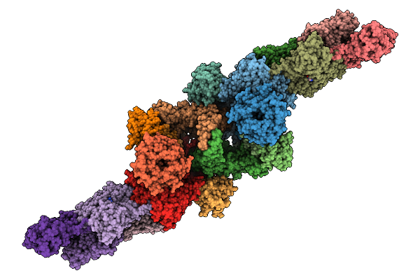
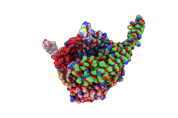
Structure Of Spin90 Dimer-Arp2/3 Complex-Nucleated Actin Filament (Singlet Complex)
Organism: Homo sapiens, Bos taurus, Gallus gallus
Method: ELECTRON MICROSCOPY Release Date: 2025-09-10 Classification: CYTOSOLIC PROTEIN Ligands: MG, ADP |
 |
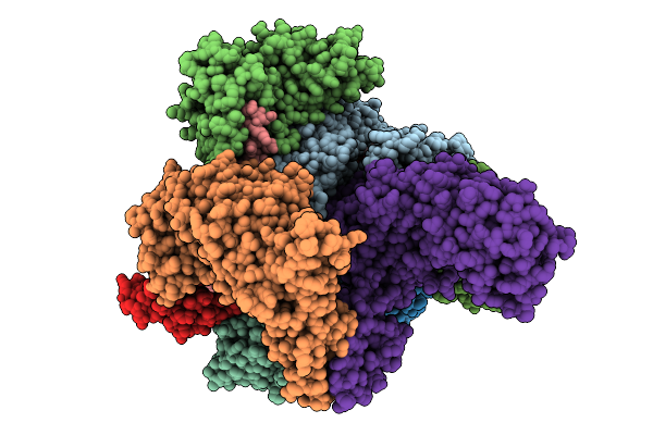
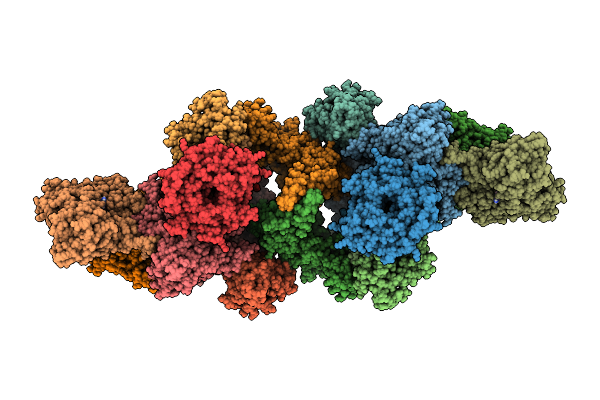
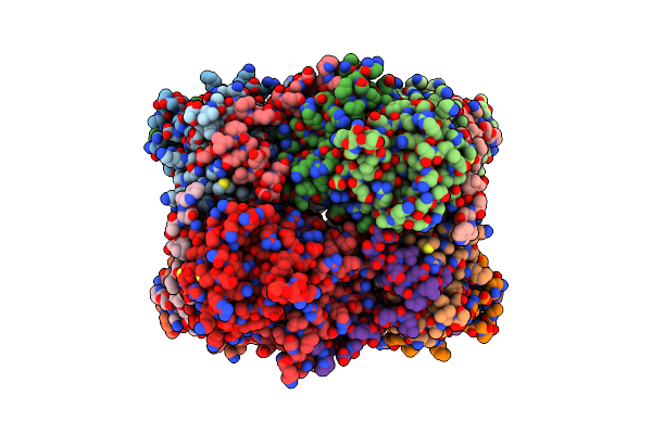
Structure Of Spin90 Dimer-Arp2/3 Complexes-Nucleated Actin Filaments (Doublet Complex)
Organism: Homo sapiens, Bos taurus, Gallus gallus
Method: ELECTRON MICROSCOPY Release Date: 2025-09-10 Classification: CYTOSOLIC PROTEIN Ligands: ADP, MG |
 |
Organism: Homo sapiens, Sus scrofa, Amanita phalloides
Method: ELECTRON MICROSCOPY Release Date: 2025-09-03 Classification: CYTOSOLIC PROTEIN Ligands: ADP, MG |
 |
Organism: Homo sapiens
Method: X-RAY DIFFRACTION Resolution:1.42 Å Release Date: 2025-08-27 Classification: SIGNALING PROTEIN |
 |
Organism: Gallus gallus
Method: X-RAY DIFFRACTION Resolution:1.34 Å Release Date: 2025-08-06 Classification: SIGNALING PROTEIN Ligands: GOL, PO4 |
 |
Organism: Gallus gallus
Method: X-RAY DIFFRACTION Resolution:1.06 Å Release Date: 2025-08-06 Classification: SIGNALING PROTEIN Ligands: GOL, PO4 |
 |
Organism: Gallus gallus
Method: X-RAY DIFFRACTION Resolution:1.13 Å Release Date: 2025-08-06 Classification: SIGNALING PROTEIN Ligands: GOL, PO4 |
 |
Organism: Gallus gallus
Method: X-RAY DIFFRACTION Resolution:1.34 Å Release Date: 2025-08-06 Classification: SIGNALING PROTEIN |
 |
Organism: Gallus gallus
Method: X-RAY DIFFRACTION Resolution:1.31 Å Release Date: 2025-08-06 Classification: STRUCTURAL PROTEIN Ligands: GOL |
 |
Organism: Gallus gallus
Method: X-RAY DIFFRACTION Resolution:1.12 Å Release Date: 2025-08-06 Classification: STRUCTURAL PROTEIN Ligands: SO4 |
 |
Organism: Gallus gallus
Method: X-RAY DIFFRACTION Resolution:1.01 Å Release Date: 2025-08-06 Classification: STRUCTURAL PROTEIN Ligands: SO4 |
 |
Organism: Homo sapiens
Method: X-RAY DIFFRACTION Resolution:2.60 Å Release Date: 2025-06-11 Classification: ENDOCYTOSIS Ligands: MES, ZN |
 |
Organism: Saccharomyces cerevisiae
Method: X-RAY DIFFRACTION Resolution:1.49 Å Release Date: 2025-05-21 Classification: ENDOCYTOSIS |
 |
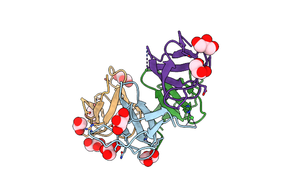
De Novo Design Of High-Affinity Protein Binders To Rna Binding Domain Of G3Bp1
Organism: Synthetic construct, Homo sapiens
Method: X-RAY DIFFRACTION Resolution:2.40 Å Release Date: 2025-05-14 Classification: DE NOVO PROTEIN |
 |
Crystal Structure Of C-Src Sh3 Domain In H32 Space Group Mediated By Nickel
Organism: Homo sapiens
Method: X-RAY DIFFRACTION Resolution:1.45 Å Release Date: 2025-05-14 Classification: SIGNALING PROTEIN Ligands: NI, GOL |
 |
Cryo-Em Structure Of The Chikv Nsp3 Peptide In Complex With The Ntf2L Domain Of G3Bp1 (Conformation I)
Organism: Homo sapiens, Chikungunya virus
Method: ELECTRON MICROSCOPY Resolution:2.66 Å Release Date: 2025-04-09 Classification: VIRAL PROTEIN |