
Search Count: 43
 |
Organism: Salmo salar
Method: X-RAY DIFFRACTION Resolution:1.89 Å Release Date: 2021-04-28 Classification: HYDROLASE |
 |
Organism: Salmo salar
Method: X-RAY DIFFRACTION Resolution:2.88 Å Release Date: 2021-04-28 Classification: HYDROLASE |
 |
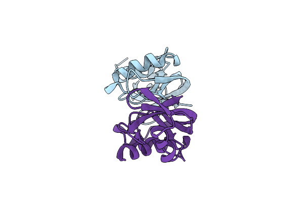
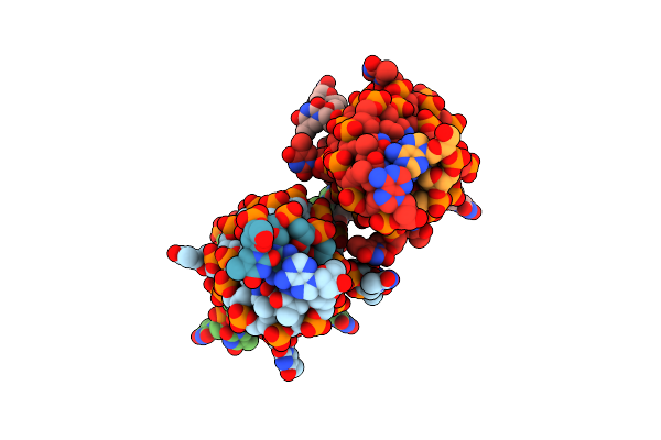
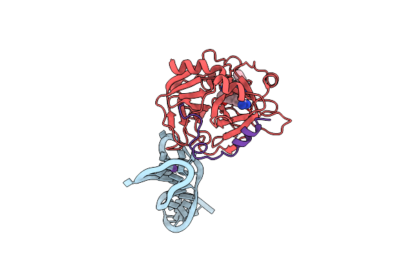
X-Ray Structure Of The Complex Between Human Alpha Thrombin And Nu172, A Duplex/Quadruplex 26-Mer Dna Aptamer, In The Presence Of Potassium Ions.
Organism: Synthetic construct, Homo sapiens
Method: X-RAY DIFFRACTION Resolution:2.50 Å Release Date: 2018-10-24 Classification: HYDROLASE Ligands: 0G6, NA, K, GOL |
 |
X-Ray Structure Of The Complex Between Human Alpha Thrombin And Nu172, A Duplex/Quadruplex 26-Mer Dna Aptamer, In The Presence Of Sodium Ions.
Organism: Synthetic construct, Homo sapiens
Method: X-RAY DIFFRACTION Resolution:2.80 Å Release Date: 2018-10-17 Classification: HYDROLASE Ligands: 0G6, NA, GOL |
 |
Organism: Thaumatococcus daniellii
Method: X-RAY DIFFRACTION Resolution:1.45 Å Release Date: 2016-12-07 Classification: PLANT PROTEIN Ligands: TLA, 73M, CPT, GOL |
 |
Organism: Homo sapiens
Method: X-RAY DIFFRACTION Resolution:2.95 Å Release Date: 2016-11-30 Classification: protein/dna Ligands: NAG, 0G6, NA |
 |
Organism: Homo sapiens
Method: X-RAY DIFFRACTION Resolution:3.59 Å Release Date: 2016-11-30 Classification: protein/dna Ligands: NAG, 0G6, NA |
 |
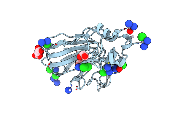
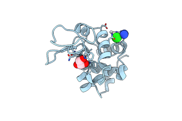
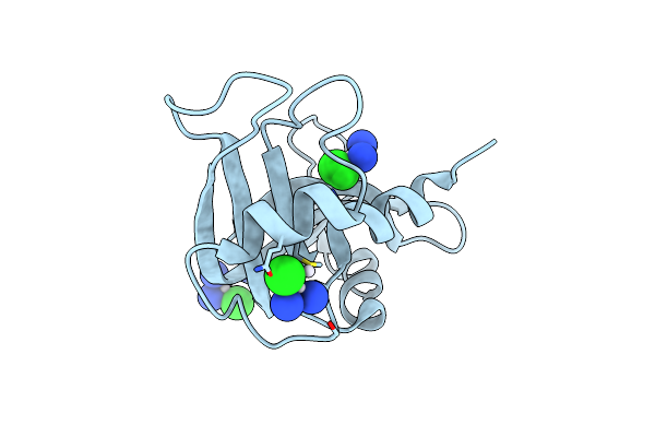
X-Ray Structure Of The Adduct Between Hen Egg White Lysozyme And Cisplatin Upon 24 Hours Of Incubation At 20 Degrees
Organism: Gallus gallus
Method: X-RAY DIFFRACTION Resolution:1.85 Å Release Date: 2016-04-13 Classification: HYDROLASE Ligands: GOL, CPT |
 |
X-Ray Structure Of The Adduct Between Hen Egg White Lysozyme And Cisplatin Upon 24 Hours Of Incubation At 55 Degrees
Organism: Gallus gallus
Method: X-RAY DIFFRACTION Resolution:1.94 Å Release Date: 2016-04-13 Classification: HYDROLASE Ligands: GOL, CPT |
 |
X-Ray Structure Of The Adduct Between Hen Egg White Lysozyme And Cisplatin At Long Incubation Times
Organism: Gallus gallus
Method: X-RAY DIFFRACTION Resolution:1.55 Å Release Date: 2016-04-13 Classification: HYDROLASE Ligands: GOL, CPT, CL |
 |
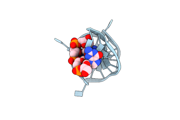
Structural Insights Into The Quadruplex-Duplex 3' Interface Formed From A Telomeric Repeat - Ttloop
Organism: Synthetic construct
Method: X-RAY DIFFRACTION Resolution:2.79 Å Release Date: 2016-01-20 Classification: DNA Ligands: K |
 |
Structural Insights Into The Quadruplex-Duplex 3' Interface Formed From A Telomeric Repeat - Tloop
Organism: Synthetic construct
Method: X-RAY DIFFRACTION Resolution:2.71 Å Release Date: 2016-01-20 Classification: DNA Ligands: K |
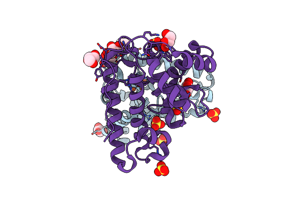 |
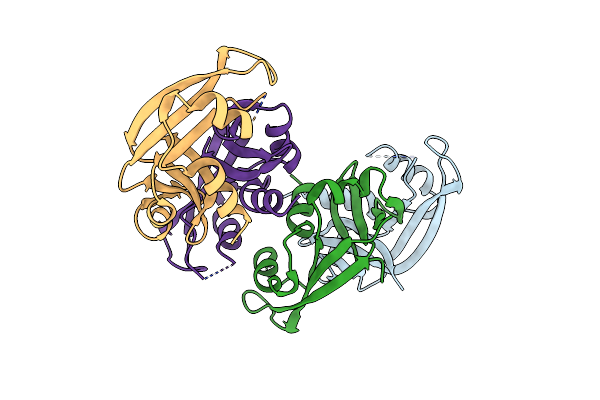
X-Ray Structure Of The Iron/Manganese Cambialistic Superoxide Dismutase From Streptococcus Thermophilus
Organism: Streptococcus thermophilus
Method: X-RAY DIFFRACTION Resolution:1.60 Å Release Date: 2016-01-13 Classification: OXIDOREDUCTASE Ligands: FE, SO4, GOL |
 |
X-Ray Structure Of The Iron/Manganese Cambialistic Superoxide Dismutase From Streptococcus Mutans
Organism: Streptococcus mutans
Method: X-RAY DIFFRACTION Resolution:2.15 Å Release Date: 2016-01-13 Classification: OXIDOREDUCTASE Ligands: FE |
 |
X-Ray Structure Of The Complex Between Human Alpha Thrombin And A Duplex/Quadruplex 31-Mer Dna Aptamer
Organism: Synthetic construct, Homo sapiens
Method: X-RAY DIFFRACTION Resolution:2.98 Å Release Date: 2016-01-13 Classification: HYDROLASE Ligands: 0G6, K |
 |
The X-Ray Structure Of Bovine Pancreatic Ribonuclease Incubated In The Presence Of An Excess Of Cisplatin (1:10 Ratio)
Organism: Bos taurus
Method: X-RAY DIFFRACTION Resolution:1.95 Å Release Date: 2015-03-25 Classification: HYDROLASE Ligands: CPT, CL |
 |
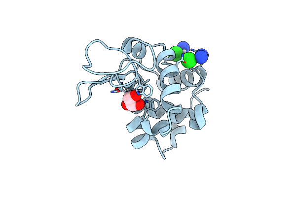
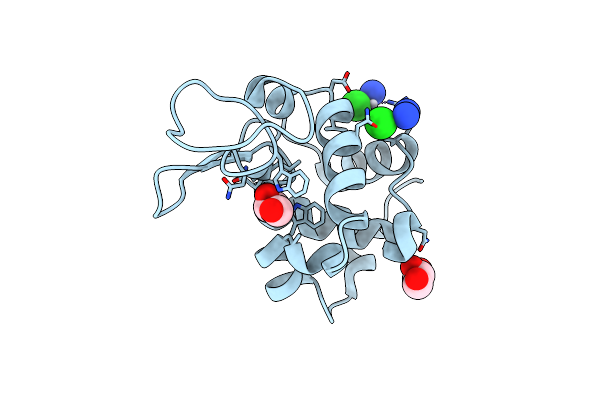
X-Ray Structure Of The Adduct Between Hen Egg White Lysozyme And Auoxo3, A Cytotoxic Gold(Iii) Compound
Organism: Gallus gallus
Method: X-RAY DIFFRACTION Resolution:2.05 Å Release Date: 2014-11-05 Classification: HYDROLASE Ligands: NO3, EDO, NA, AU |
 |
X-Ray Structure Of The Adduct Formed Between Hen Egg White Lysozyme And Trans-Dimethylamine Methylamine Dichlorido Platinum(Ii)
Organism: Gallus gallus
Method: X-RAY DIFFRACTION Resolution:2.51 Å Release Date: 2014-07-30 Classification: HYDROLASE Ligands: I83 |
 |
X-Ray Structure Of The Adduct Formed Between Bovine Pancreatic Ribonuclease And Trans-Dimethylamine Methylamine Dichlorido Platinum(Ii)
Organism: Bos taurus
Method: X-RAY DIFFRACTION Resolution:2.00 Å Release Date: 2014-07-30 Classification: HYDROLASE Ligands: I83 |
 |
Method: X-RAY DIFFRACTION
Resolution:1.70 Å Release Date: 2014-03-05 Classification: DNA, RNA Ligands: K, GOL |