
Search Count: 24
 |
Organism: Methanosarcina mazei go1, Synthetic construct
Method: X-RAY DIFFRACTION Resolution:2.11 Å Release Date: 2023-11-22 Classification: LYASE Ligands: SO4, FDA |
 |
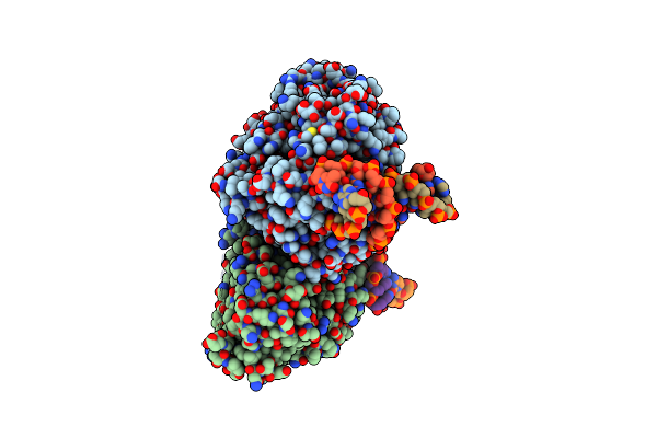
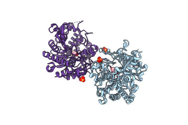
Time-Resolved Sfx Structure Of The Class Ii Photolyase Complexed With A Thymine Dimer (3 Picosecond Pump-Probe Delay)
Organism: Methanosarcina mazei go1, Synthetic construct
Method: X-RAY DIFFRACTION Resolution:2.16 Å Release Date: 2023-11-22 Classification: LYASE Ligands: SO4, FDA |
 |
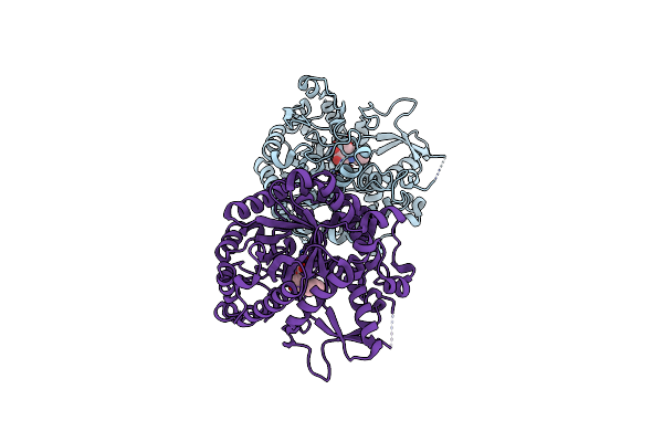
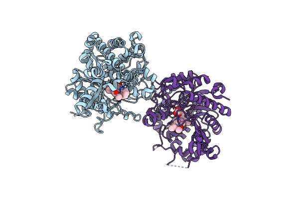
Time-Resolved Sfx Structure Of The Class Ii Photolyase Complexed With A Thymine Dimer (300 Ps Pump-Probe Delay)
Organism: Methanosarcina mazei go1, Synthetic construct
Method: X-RAY DIFFRACTION Resolution:2.35 Å Release Date: 2023-11-22 Classification: LYASE Ligands: SO4, FDA |
 |
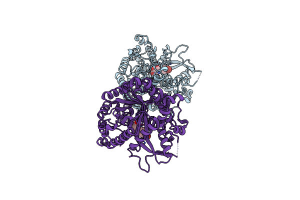
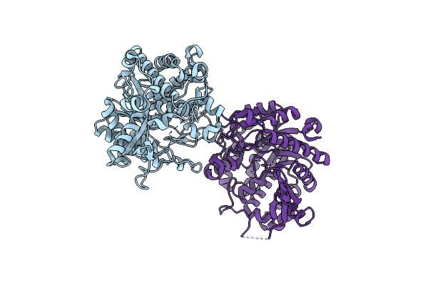
Time-Resolved Sfx Structure Of The Class Ii Photolyase Complexed With A Thymine Dimer (1 Nanosecond Pump-Probe Delay)
Organism: Methanosarcina mazei go1, Synthetic construct
Method: X-RAY DIFFRACTION Resolution:2.27 Å Release Date: 2023-11-22 Classification: LYASE Ligands: SO4, FDA |
 |
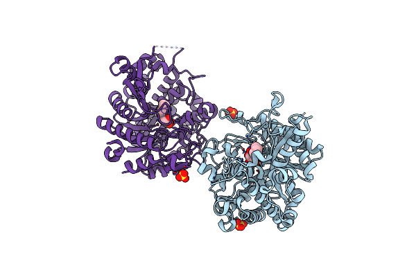
Time-Resolved Sfx Structure Of The Class Ii Photolyase Complexed With A Thymine Dimer (3 Nanosecond Pump-Probe Delay)
Organism: Methanosarcina mazei go1, Synthetic construct
Method: X-RAY DIFFRACTION Resolution:2.35 Å Release Date: 2023-11-22 Classification: LYASE Ligands: FDA, SO4 |
 |
Time-Resolved Sfx Structure Of The Class Ii Photolyase Complexed With A Thymine Dimer (10 Nanosecond Pump-Probe Delay)
Organism: Methanosarcina mazei go1, Synthetic construct
Method: X-RAY DIFFRACTION Resolution:2.36 Å Release Date: 2023-11-22 Classification: LYASE Ligands: FDA, SO4 |
 |
Time-Resolved Sfx Structure Of The Class Ii Photolyase Complexed With A Thymine Dimer (30 Nanosecond Timepoint)
Organism: Methanosarcina mazei go1, Synthetic construct
Method: X-RAY DIFFRACTION Resolution:2.39 Å Release Date: 2023-11-22 Classification: LYASE Ligands: SO4, FDA |
 |
Time-Resolved Sfx Structure Of The Class Ii Photolyase Complexed With A Thymine Dimer (1 Microsecond Pump-Probe Delay)
Organism: Methanosarcina mazei go1, Synthetic construct
Method: X-RAY DIFFRACTION Resolution:2.24 Å Release Date: 2023-11-22 Classification: LYASE Ligands: SO4, FDA |
 |
Time-Resolved Sfx Structure Of The Class Ii Photolyase Complexed With A Thymine Dimer (10 Microsecond Pump Probe Delay)
Organism: Methanosarcina mazei go1, Synthetic construct
Method: X-RAY DIFFRACTION Resolution:2.18 Å Release Date: 2023-11-22 Classification: LYASE Ligands: SO4, FDA |
 |
Time-Resolved Sfx Structure Of The Class Ii Photolyase Complexed With A Thymine Dimer (30 Microsecond Pump-Probe Delay)
Organism: Methanosarcina mazei go1, Synthetic construct
Method: X-RAY DIFFRACTION Resolution:2.25 Å Release Date: 2023-11-22 Classification: LYASE Ligands: SO4, FDA |
 |
Time-Resolved Sfx Structure Of The Class Ii Photolyase Complexed With A Thymine Dimer (100 Microsecond Timpeoint)
Organism: Methanosarcina mazei go1, Synthetic construct
Method: X-RAY DIFFRACTION Resolution:2.50 Å Release Date: 2023-11-22 Classification: LYASE Ligands: SO4, FDA |
 |
Organism: Rauvolfia serpentina
Method: X-RAY DIFFRACTION Resolution:2.50 Å Release Date: 2014-02-05 Classification: HYDROLASE Ligands: C1K |
 |
Organism: Rauvolfia serpentina
Method: X-RAY DIFFRACTION Resolution:3.01 Å Release Date: 2014-02-05 Classification: HYDROLASE Ligands: LR1 |
 |
Organism: Rauvolfia serpentina
Method: X-RAY DIFFRACTION Resolution:2.40 Å Release Date: 2014-01-29 Classification: HYDROLASE Ligands: SO4, VM2 |
 |
Organism: Rauvolfia serpentina
Method: X-RAY DIFFRACTION Resolution:2.30 Å Release Date: 2013-02-20 Classification: HYDROLASE Ligands: BGC, CL |
 |
Organism: Rauvolfia serpentina
Method: X-RAY DIFFRACTION Resolution:2.52 Å Release Date: 2013-01-30 Classification: HYDROLASE Ligands: BGC |
 |
Organism: Rauvolfia serpentina
Method: X-RAY DIFFRACTION Resolution:2.10 Å Release Date: 2013-01-16 Classification: HYDROLASE Ligands: SO4 |
 |
Crystal Structure Of Raucaffricine Glucosidase From Ajmaline Biosynthesis Pathway
Organism: Rauvolfia serpentina
Method: X-RAY DIFFRACTION Resolution:2.15 Å Release Date: 2012-08-15 Classification: HYDROLASE Ligands: SO4, GOL |
 |
Structures Of Alkaloid Biosynthetic Glucosidases Decode Substrate Specificity
Organism: Rauvolfia serpentina
Method: X-RAY DIFFRACTION Resolution:2.43 Å Release Date: 2011-11-30 Classification: HYDROLASE Ligands: DH8, CL |
 |
Structures Of Alkaloid Biosynthetic Glucosidases Decode Substrate Specificity
Organism: Rauvolfia serpentina
Method: X-RAY DIFFRACTION Resolution:2.20 Å Release Date: 2011-11-30 Classification: HYDROLASE Ligands: CL |