
Search Count: 40
 |
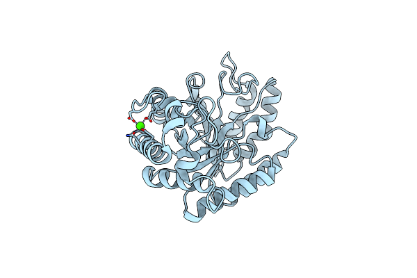
Organism: Streptomyces sp. f-1
Method: X-RAY DIFFRACTION Resolution:1.55 Å Release Date: 2019-12-04 Classification: ISOMERASE Ligands: SO4, MG |
 |
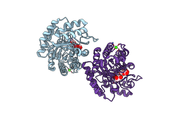
Organism: Streptomyces sp. f-1
Method: X-RAY DIFFRACTION Resolution:2.80 Å Release Date: 2019-12-04 Classification: ISOMERASE Ligands: MG, SO4, MPD |
 |
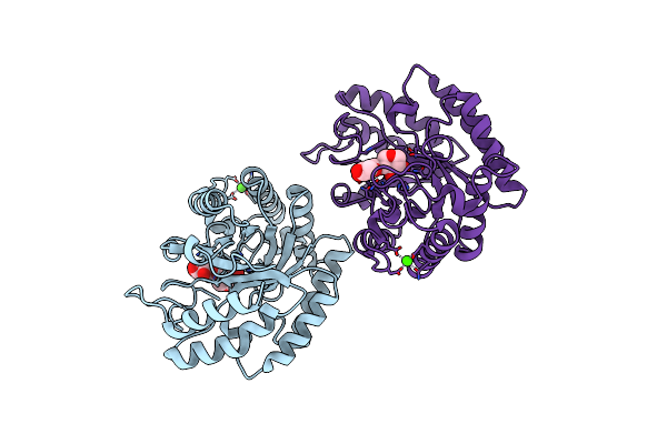
Organism: Bacillus licheniformis (strain atcc 14580 / dsm 13 / jcm 2505 / nbrc 12200 / ncimb 9375 / nrrl nrs-1264 / gibson 46)
Method: X-RAY DIFFRACTION Resolution:2.49 Å Release Date: 2019-04-17 Classification: HYDROLASE Ligands: CA |
 |
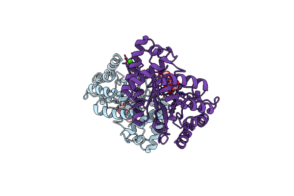
Crystal Structure Of The Gh43 Protein Blxynb Mutant (K247S) From Bacillus Licheniformis
Organism: Bacillus licheniformis (strain atcc 14580 / dsm 13 / jcm 2505 / nbrc 12200 / ncimb 9375 / nrrl nrs-1264 / gibson 46)
Method: X-RAY DIFFRACTION Resolution:1.95 Å Release Date: 2019-04-17 Classification: HYDROLASE Ligands: CA, SO4, GOL, IMD |
 |
Crystal Structure Of A Gh1 Beta-Glucosidase Retrieved From Microbial Metagenome Of Poraque Amazon Lake
Organism: Metagenome
Method: X-RAY DIFFRACTION Resolution:2.75 Å Release Date: 2018-03-07 Classification: HYDROLASE Ligands: GOL, PEG |
 |
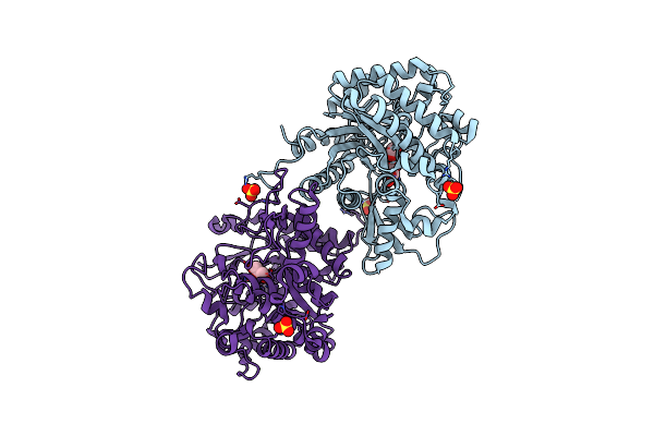
Organism: Coptotermes gestroi
Method: X-RAY DIFFRACTION Resolution:2.85 Å Release Date: 2017-01-18 Classification: OXIDOREDUCTASE Ligands: NAP |
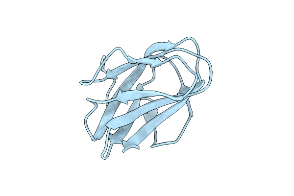 |
Structure Of Cbm_E1, A Novel Carbohydrate-Binding Module Found By Sugar Cane Soil Metagenome
Organism: Uncultured bacterium
Method: X-RAY DIFFRACTION Resolution:1.75 Å Release Date: 2016-09-21 Classification: SUGAR BINDING PROTEIN |
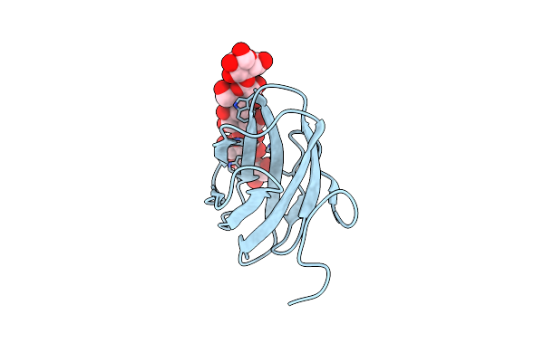 |
Structure Of Cbm_E1, A Novel Carbohydrate-Binding Module Found By Sugar Cane Soil Metagenome, Complexed With Cellopentaose
Organism: Uncultured bacterium
Method: X-RAY DIFFRACTION Resolution:1.50 Å Release Date: 2016-09-21 Classification: SUGAR BINDING PROTEIN |
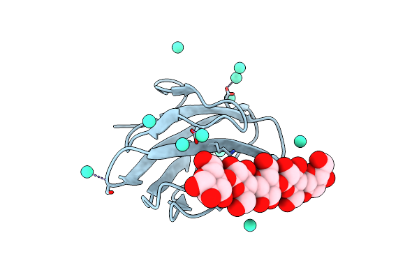 |
Structure Of Cbm_E1, A Novel Carbohydrate-Binding Module Found By Sugar Cane Soil Metagenome, Complexed With Cellopentaose And Gadolinium Ion
Organism: Uncultured bacterium
Method: X-RAY DIFFRACTION Resolution:1.80 Å Release Date: 2016-09-21 Classification: SUGAR BINDING PROTEIN Ligands: GD |
 |
Organism: Trichoderma harzianum
Method: X-RAY DIFFRACTION Resolution:2.60 Å Release Date: 2016-07-06 Classification: HYDROLASE Ligands: GOL, SO4 |
 |
Crystal Structure Of The Gh1 Beta-Glucosidase From Exiguobacterium Antarcticum B7 In Space Group P21
Organism: Exiguobacterium antarcticum (strain b7)
Method: X-RAY DIFFRACTION Resolution:2.24 Å Release Date: 2016-04-13 Classification: HYDROLASE Ligands: SO4 |
 |
Crystal Structure Of The Gh1 Beta-Glucosidase From Exiguobacterium Antarcticum B7 In Space Group C2221
Organism: Exiguobacterium antarcticum (strain b7)
Method: X-RAY DIFFRACTION Resolution:2.15 Å Release Date: 2016-04-13 Classification: HYDROLASE Ligands: CXS, SO4, GOL |
 |
Crystal Structure Of A Novel Reducing-End Xylose-Releasing Exo-Oligoxylanase (Xyna) Belonging To Gh10 Family (Space Group P1211)
Organism: Xanthomonas axonopodis pv. citri
Method: X-RAY DIFFRACTION Resolution:2.86 Å Release Date: 2014-10-08 Classification: HYDROLASE |
 |
Crystal Structure Of A Novel Reducing-End Xylose-Releasing Exo-Oligoxylanase (Xyna) Belonging To Gh10 Family (Space Group P43212)
Organism: Xanthomonas axonopodis pv. citri
Method: X-RAY DIFFRACTION Resolution:3.00 Å Release Date: 2014-10-08 Classification: HYDROLASE |
 |
Crystal Structure Of Gh10 Endo-B-1,4-Xylanase (Xynb) From Xanthomonas Axonopodis Pv Citri In The Native Form
Organism: Xanthomonas axonopodis pv. citri
Method: X-RAY DIFFRACTION Resolution:1.30 Å Release Date: 2014-10-08 Classification: HYDROLASE Ligands: CA |
 |
Crystal Structure Of Gh10 Endo-B-1,4-Xylanase (Xynb) From Xanthomonas Axonopodis Pv Citri Complexed With Xylose
Organism: Xanthomonas axonopodis pv. citri
Method: X-RAY DIFFRACTION Resolution:1.60 Å Release Date: 2014-10-08 Classification: HYDROLASE Ligands: XYP, GOL, CA |
 |
Crystal Structure Of Gh10 Endo-B-1,4-Xylanase (Xynb) From Xanthomonas Axonopodis Pv Citri Complexed With Xylobiose
Organism: Xanthomonas axonopodis pv. citri
Method: X-RAY DIFFRACTION Resolution:1.40 Å Release Date: 2014-10-08 Classification: HYDROLASE Ligands: CA |
 |
Crystal Structure Of Gh10 Endo-B-1,4-Xylanase (Xynb) From Xanthomonas Axonopodis Pv Citri Complexed With Xylotriose
Organism: Xanthomonas axonopodis pv. citri
Method: X-RAY DIFFRACTION Resolution:1.42 Å Release Date: 2014-10-08 Classification: HYDROLASE Ligands: CA, XYP |
 |
Crystal Structure Of Endo-1,5-Alpha-L-Arabinanase From Thermotoga Petrophila Rku-1
Organism: Thermotoga petrophila
Method: X-RAY DIFFRACTION Resolution:1.75 Å Release Date: 2014-02-05 Classification: HYDROLASE Ligands: PEG, CA |
 |
Crystal Structure Of Endo-1,5-Alpha-L-Arabinanase From Thermotoga Petrophila Rku-1 In Complex With Tris
Organism: Thermotoga petrophila
Method: X-RAY DIFFRACTION Resolution:1.76 Å Release Date: 2014-02-05 Classification: HYDROLASE Ligands: TRS, CA |