
Search Count: 14
 |
Organism: Severe acute respiratory syndrome coronavirus 2, Vicugna pacos
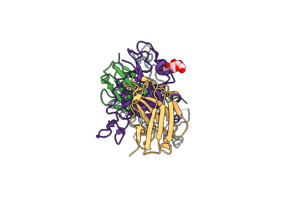
Method: X-RAY DIFFRACTION Resolution:2.90 Å Release Date: 2021-05-12 Classification: VIRAL PROTEIN Ligands: NAG |
 |
Organism: Severe acute respiratory syndrome coronavirus 2, Vicugna pacos
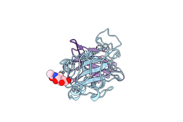
Method: X-RAY DIFFRACTION Resolution:2.30 Å Release Date: 2021-05-12 Classification: VIRAL PROTEIN Ligands: NAG, PO4, PEG |
 |
Organism: Severe acute respiratory syndrome coronavirus 2, Camelus bactrianus
Method: ELECTRON MICROSCOPY Release Date: 2021-04-28 Classification: VIRAL PROTEIN Ligands: NAG |
 |
Organism: Severe acute respiratory syndrome coronavirus 2, Lama glama, Vicugna pacos
Method: ELECTRON MICROSCOPY Release Date: 2021-04-28 Classification: VIRAL PROTEIN Ligands: NAG |
 |
Organism: Severe acute respiratory syndrome coronavirus 2, Vicugna pacos, Lama glama
Method: ELECTRON MICROSCOPY Release Date: 2021-02-10 Classification: VIRAL PROTEIN Ligands: NAG |
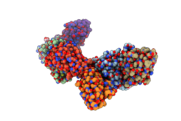 |
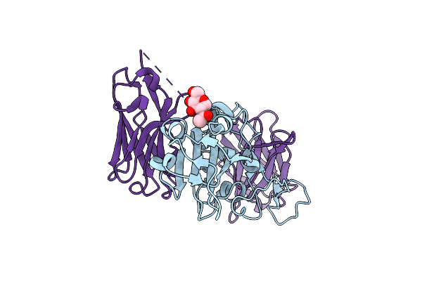
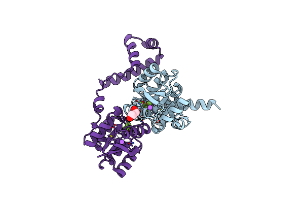
Crystal Structure Of Sars-Cov-2 Receptor Binding Domain Complexed With Nanobodies Vhh E And U
Organism: Severe acute respiratory syndrome coronavirus 2, Vicugna pacos
Method: X-RAY DIFFRACTION Resolution:1.87 Å Release Date: 2021-01-20 Classification: VIRAL PROTEIN/IMMUNE SYSTEM Ligands: EDO, NAG |
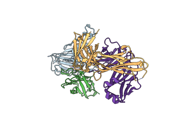 |
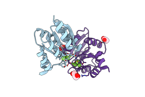
Crystal Structure Of Sars-Cov-2 Receptor Binding Domain Complexed With Nanobody Vhh V And Antibody Fab Cc12.3
Organism: Severe acute respiratory syndrome coronavirus 2, Vicugna pacos, Homo sapiens
Method: X-RAY DIFFRACTION Resolution:2.55 Å Release Date: 2021-01-20 Classification: VIRAL PROTEIN/IMMUNE SYSTEM Ligands: NAG |
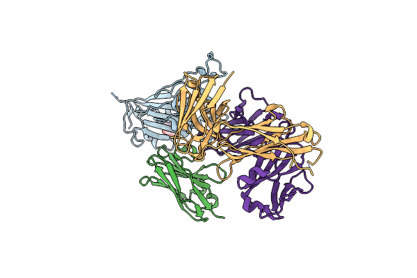 |
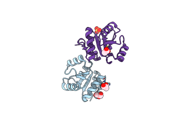
Crystal Structure Of Sars-Cov-2 Receptor Binding Domain Complexed With Nanobody Vhh W And Antibody Fab Cc12.3
Organism: Severe acute respiratory syndrome coronavirus 2, Vicugna pacos, Homo sapiens
Method: X-RAY DIFFRACTION Resolution:2.73 Å Release Date: 2021-01-20 Classification: VIRAL PROTEIN/IMMUNE SYSTEM Ligands: NAG |
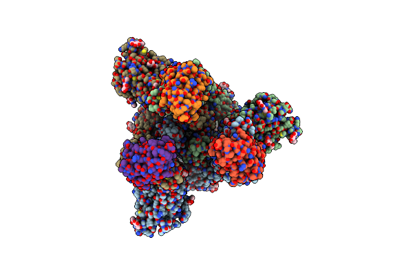 |
Organism: Severe acute respiratory syndrome coronavirus 2, Lama glama
Method: ELECTRON MICROSCOPY Release Date: 2021-01-20 Classification: VIRAL PROTEIN/IMMUNE SYSTEM Ligands: NAG |
 |
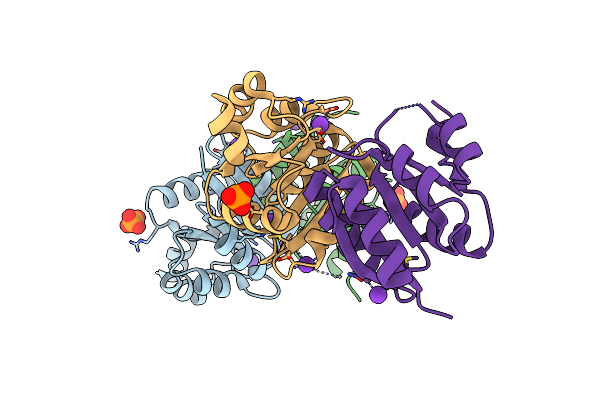
Crystal Structure Of The Full-Length Response Regulator Desr In The Active State
Organism: Bacillus subtilis subsp. subtilis
Method: X-RAY DIFFRACTION Resolution:2.31 Å Release Date: 2014-07-16 Classification: DNA BINDING PROTEIN Ligands: MG, BEF, GOL, NA |
 |
Crystal Structure Of The Receiver Domain Of Desr In Complex With Beryllofluoride And Magnesium
Organism: Bacillus subtilis subsp. subtilis
Method: X-RAY DIFFRACTION Resolution:2.27 Å Release Date: 2014-07-16 Classification: DNA BINDING PROTEIN Ligands: MG, BEF, ACT, GOL |
 |
Organism: Bacillus subtilis subsp. subtilis
Method: X-RAY DIFFRACTION Resolution:1.95 Å Release Date: 2014-07-16 Classification: DNA BINDING PROTEIN Ligands: GOL, SO4, ACT |
 |
Crystal Structure Of The Unphosphorylated Receiver Domain Of Desr In The Active State
Organism: Bacillus subtilis subsp. subtilis
Method: X-RAY DIFFRACTION Resolution:2.54 Å Release Date: 2014-07-16 Classification: DNA BINDING PROTEIN Ligands: PO4, K |
 |
Organism: Bacillus subtilis
Method: X-RAY DIFFRACTION Resolution:1.74 Å Release Date: 2009-09-15 Classification: TRANSFERASE Ligands: ATP, MG, IOD |