
Search Count: 13
All
Selected
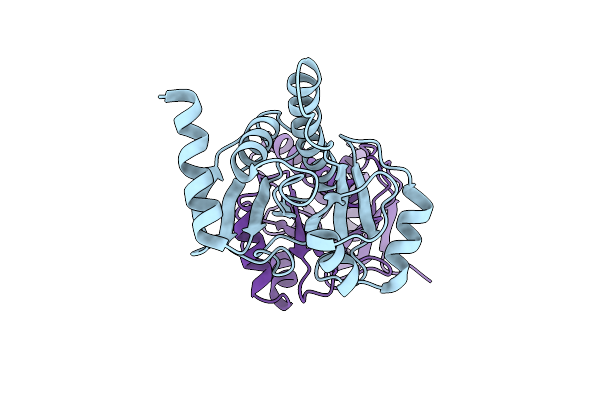 |
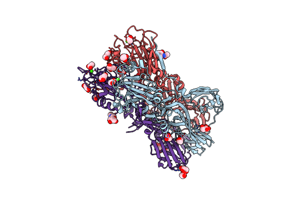
Organism: Rubella virus
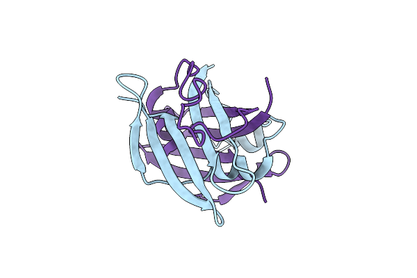
Method: X-RAY DIFFRACTION Resolution:1.72 Å Release Date: 2024-01-17 Classification: HYDROLASE |
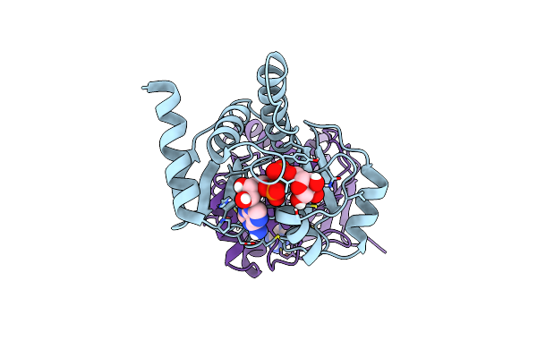 |
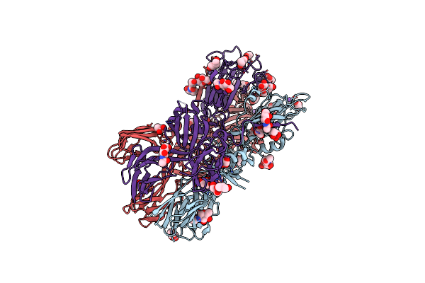
Organism: Rubella virus
Method: X-RAY DIFFRACTION Resolution:1.59 Å Release Date: 2024-01-17 Classification: HYDROLASE Ligands: APR |
 |
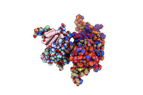
Organism: Rubella virus (strain ra27/3 vaccine)
Method: X-RAY DIFFRACTION Resolution:1.64 Å Release Date: 2022-07-13 Classification: HYDROLASE Ligands: GOL, EDO, PEG, ZN |
 |
Structure Of Rubella Virus E1 Glycoprotein Ectodomain Fitted Into Sub-Tomogram Averaged Surface Spike Density Of Rubella Virus
Organism: Rubella virus
Method: ELECTRON MICROSCOPY Release Date: 2017-05-10 Classification: VIRAL PROTEIN |
 |
Organism: Rubella virus
Method: ELECTRON MICROSCOPY Release Date: 2017-05-10 Classification: VIRAL PROTEIN |
 |
Organism: Rubella virus
Method: ELECTRON MICROSCOPY Release Date: 2017-05-10 Classification: VIRAL PROTEIN |
 |
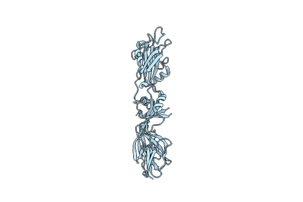
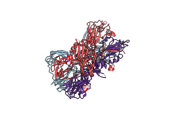
Organism: Rubella virus
Method: X-RAY DIFFRACTION Resolution:2.66 Å Release Date: 2013-12-11 Classification: VIRAL PROTEIN Ligands: LDA, CL |
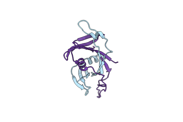 |
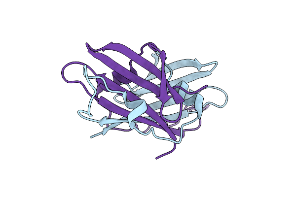
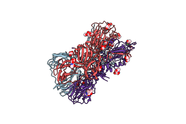
Organism: Rubella virus
Method: X-RAY DIFFRACTION Resolution:2.30 Å Release Date: 2013-12-11 Classification: VIRAL PROTEIN |
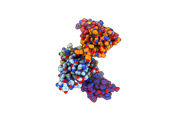 |
Organism: Rubella virus
Method: X-RAY DIFFRACTION Resolution:3.24 Å Release Date: 2013-12-11 Classification: VIRAL PROTEIN |
 |
Crystal Structure Of The Rubella Virus Envelope Glycoprotein E1 In Post-Fusion Form (Crystal Form Ii)
Organism: Rubella virus
Method: X-RAY DIFFRACTION Resolution:2.18 Å Release Date: 2013-01-09 Classification: VIRAL PROTEIN Ligands: NAG, GOL, CA, ACT, CL, NGA, PEG |
 |
Crystal Structure Of The Rubella Virus Envelope Glycoprotein E1 In Post-Fusion Form (Crystal Form I)
Organism: Rubella virus
Method: X-RAY DIFFRACTION Resolution:1.80 Å Release Date: 2013-01-09 Classification: VIRAL PROTEIN Ligands: NAG, GOL, NA, ACT, PEG, NGA |
 |
Crystal Structure Of The Rubella Virus Glycoprotein E1 In Its Post-Fusion Form Crystallized In Presence Of 1Mm Of Calcium Acetate
Organism: Rubella virus
Method: X-RAY DIFFRACTION Resolution:1.94 Å Release Date: 2013-01-09 Classification: VIRAL PROTEIN Ligands: NAG, GOL, NA, CA, ACT |
 |
Crystal Structure Of The Rubella Virus Glycoprotein E1 In Its Post-Fusion Form Crystallized In Presence Of 20Mm Of Calcium Acetate
Organism: Rubella virus
Method: X-RAY DIFFRACTION Resolution:1.98 Å Release Date: 2013-01-09 Classification: VIRAL PROTEIN Ligands: NAG, NGA, GOL, ACT, PG4, PEG, NA, CA |