
Search Count: 13
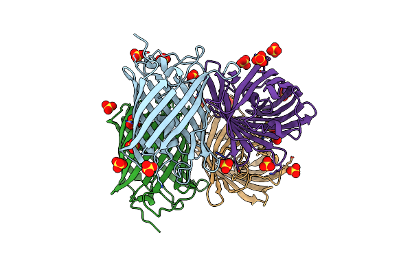 |
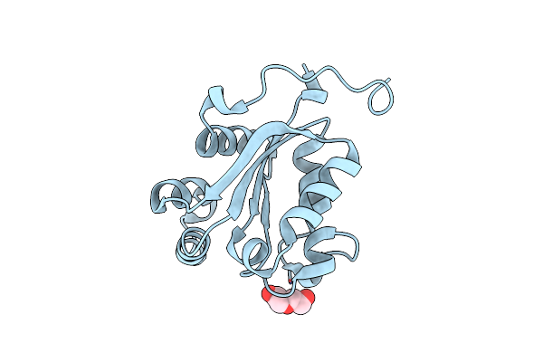
Organism: Lobophyllia hemprichii
Method: X-RAY DIFFRACTION Resolution:1.90 Å Release Date: 2017-01-11 Classification: FLUORESCENT PROTEIN Ligands: SO4 |
 |
Organism: Streptomyces lividans
Method: X-RAY DIFFRACTION Resolution:1.31 Å Release Date: 2016-09-07 Classification: HYDROLASE |
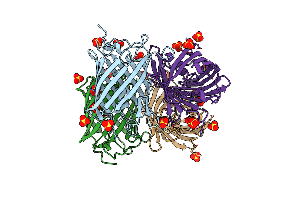 |
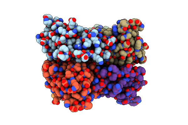
Organism: Lobophyllia hemprichii
Method: X-RAY DIFFRACTION Resolution:2.40 Å Release Date: 2016-04-13 Classification: FLUORESCENT PROTEIN Ligands: SO4 |
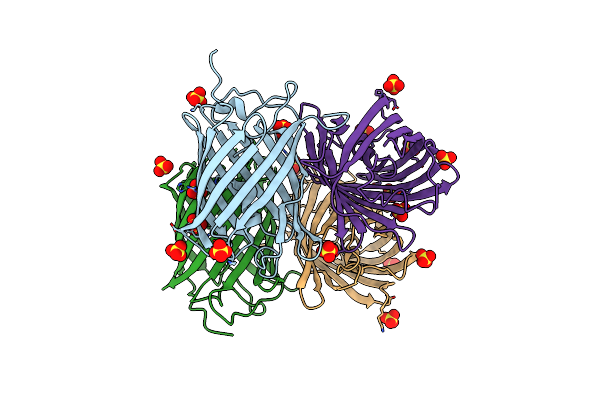 |
Room Temperature Structure Of Irisfp Determined By Serial Femtosecond Crystallography.
Organism: Lobophyllia hemprichii
Method: X-RAY DIFFRACTION Resolution:2.75 Å Release Date: 2016-04-06 Classification: FLUORESCENT PROTEIN Ligands: SO4, NH4 |
 |
Organism: Dictyostelium discoideum
Method: X-RAY DIFFRACTION Resolution:2.32 Å Release Date: 2015-02-11 Classification: TRANSFERASE Ligands: EOI |
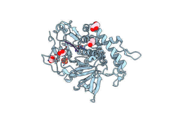 |
Crystal Structure Of The Catalytic Domain Of Botulinum Neurotoxin Bont/A C134S Mutant With Covalent Inhibitor That Modifies Cys-165 Causing Disorder In 167-174 Stretch
Organism: Clostridium botulinum a, Synthetic construct
Method: X-RAY DIFFRACTION Resolution:2.59 Å Release Date: 2014-10-15 Classification: HYDROLASE/HYDROLASE INHIBITOR Ligands: ZN, PGO, SO4, GOL |
 |
Crystal Structure Of The Catalytic Domain Of Botulinum Neurotoxin Bont/A C134 Mutant With Mtsea Modified Cys-165 Causing Stretch Disorder
Organism: Clostridium botulinum a
Method: X-RAY DIFFRACTION Resolution:1.70 Å Release Date: 2014-07-09 Classification: HYDROLASE Ligands: ZN, DMS, LMR, GOL, EDO, NA |
 |
Crystal Structure Of The Catalytic Domain Of Botulinum Neurotoxin Bont/A C134S Mutant With Covalent Inhibitor That Modifies Cys-165 Causing Disorder In 166-174 Stretch
Organism: Clostridium botulinum a, Synthetic construct
Method: X-RAY DIFFRACTION Resolution:1.93 Å Release Date: 2014-06-25 Classification: HYDROLASE/HYDROLASE INHIBITOR Ligands: ZN, SO4, GOL, PEG, PGO, EDO |
 |
Organism: Streptococcus pneumoniae
Method: SOLUTION NMR Release Date: 2013-11-20 Classification: ZINC-BINDING PROTEIN Ligands: ZN |
 |
Organism: Dictyostelium discoideum
Method: X-RAY DIFFRACTION Resolution:1.25 Å Release Date: 2013-11-20 Classification: TRANSFERASE Ligands: PEG |
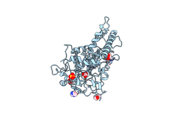 |
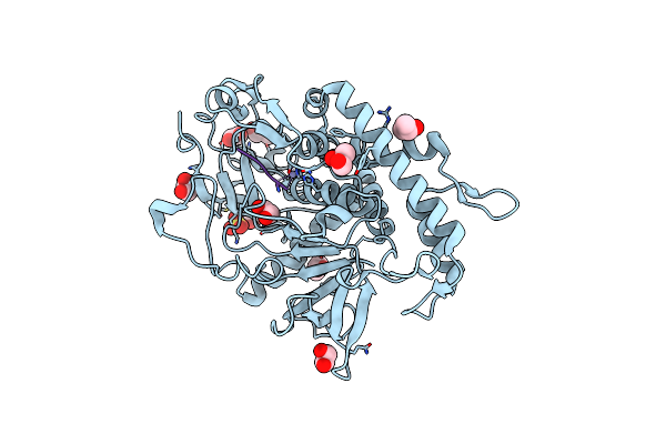
Crystal Structure Of The Catalytic Domain Of Botulinum Neurotoxin Bont/A Wild-Type
Organism: Clostridium botulinum
Method: X-RAY DIFFRACTION Resolution:1.87 Å Release Date: 2012-08-15 Classification: HYDROLASE Ligands: ZN, EDO, SO4, IMD, CO3, GOL |
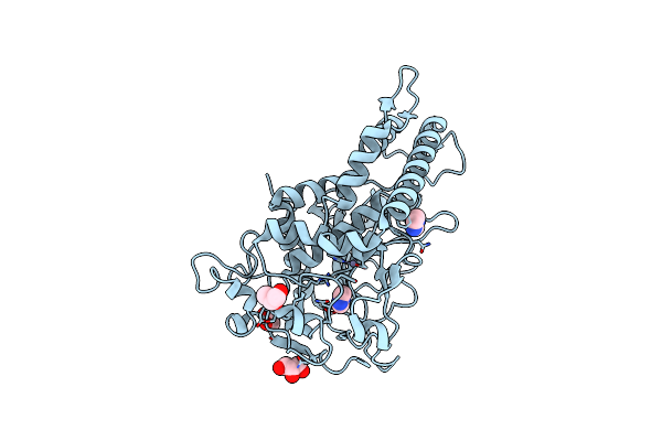 |
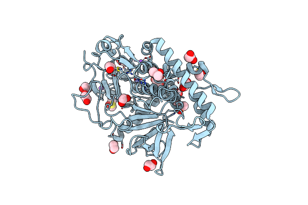
Crystal Structure Of The Catalytic Domain Of Botulinum Neurotoxin Bont/A C134S/C165S Double Mutant
Organism: Clostridium botulinum
Method: X-RAY DIFFRACTION Resolution:1.20 Å Release Date: 2012-08-15 Classification: HYDROLASE Ligands: ZN, GOL, IMD, EDO |
 |
Crystal Structure Of The Catalytic Domain Of Botulinum Neurotoxin Bont/A C134 Mutant With Mtsea Modified Cys-165
Organism: Clostridium botulinum
Method: X-RAY DIFFRACTION Resolution:1.80 Å Release Date: 2012-08-15 Classification: HYDROLASE Ligands: ZN, IMD, LMR, DMS, SO4, MPD, LI |