
Search Count: 14
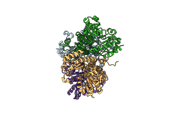 |
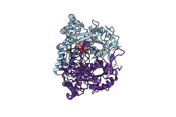
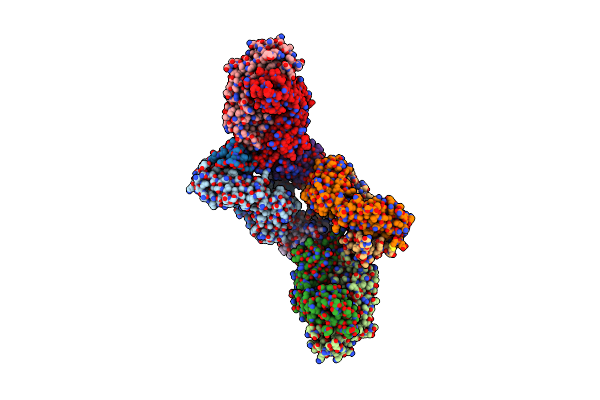
Crystal Structure Of Pseudomonas Putida Methionine Gamma-Lyase Q349S Mutant Ligand-Free Form.
Organism: Pseudomonas putida
Method: X-RAY DIFFRACTION Resolution:2.40 Å Release Date: 2022-04-20 Classification: LYASE |
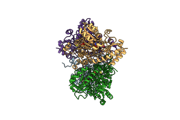 |
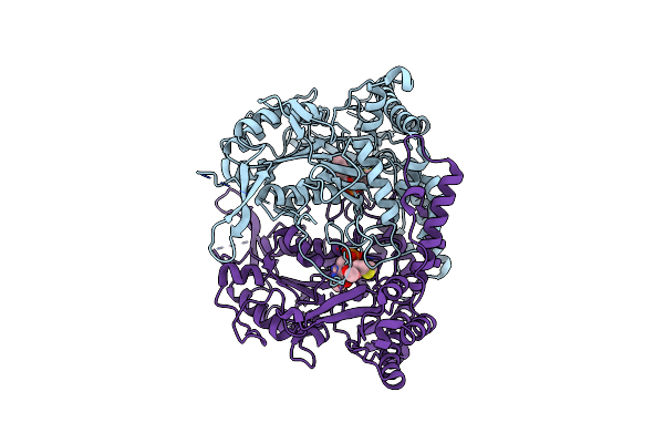
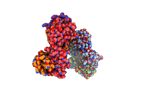
Crystal Structure Of Pseudomonas Putida Methionine Gamma-Lyase Q349S Mutant With L-Methionine Intermediates
Organism: Pseudomonas putida
Method: X-RAY DIFFRACTION Resolution:2.40 Å Release Date: 2022-04-20 Classification: LYASE Ligands: 3LM, MET |
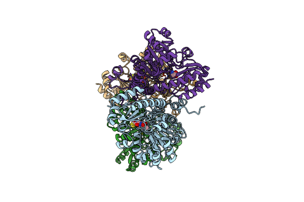 |
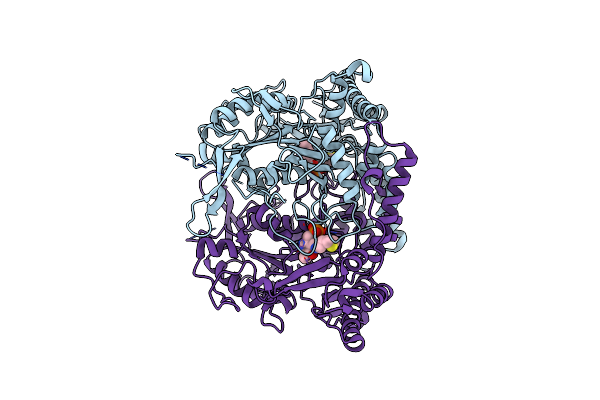
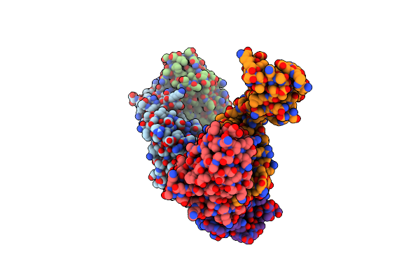
Crystal Structure Of Pseudomonas Putida Methionine Gamma-Lyase Q349S Mutant With L-Homocysteine Intermediates
Organism: Pseudomonas putida
Method: X-RAY DIFFRACTION Release Date: 2022-04-20 Classification: LYASE Ligands: 7XF, HCS |
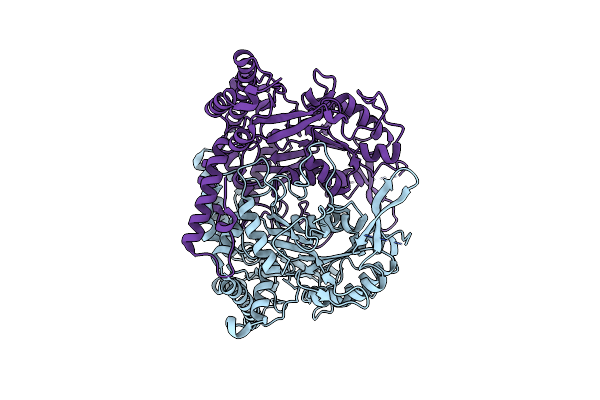 |
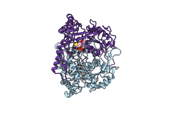
Crystal Structure Of L-Methionine Decarboxylase From Streptomyces Sp.590 (Internal Aldimine Form).
Organism: Streptomyces sp. 590 ki-2014
Method: X-RAY DIFFRACTION Resolution:1.80 Å Release Date: 2021-01-27 Classification: LYASE |
 |
Crystal Structure Of L-Methionine Decarboxylase Q64A Mutant From Streptomyces Sp.590 In Complexed With L- Methionine Methyl Ester (Geminal Diamine Form).
Organism: Streptomyces sp. 590 ki-2014
Method: X-RAY DIFFRACTION Resolution:1.45 Å Release Date: 2021-01-27 Classification: LYASE Ligands: G03 |
 |
Crystal Structure Of L-Methionine Decarboxylase From Streptomyces Sp.590 In Complexed With L- Methionine Methyl Ester (External Aldimine Form).
Organism: Streptomyces sp. 590 ki-2014
Method: X-RAY DIFFRACTION Resolution:1.51 Å Release Date: 2021-01-27 Classification: LYASE Ligands: G06 |
 |
Crystal Structure Of L-Methionine Decarboxylase From Streptomyces Sp.590 In Complexed With 3-Methlythiopropylamine (External Aldimine Form).
Organism: Streptomyces sp. 590 ki-2014
Method: X-RAY DIFFRACTION Resolution:1.61 Å Release Date: 2021-01-27 Classification: LYASE Ligands: G0C |
 |
Crystal Structure Of L-Methionine Decarboxylase From Streptomyces Sp.590 In Complexed With 3-Methlythiopropylamine (Geminal Diamine Form).
Organism: Streptomyces sp. 590 ki-2014
Method: X-RAY DIFFRACTION Resolution:1.80 Å Release Date: 2021-01-27 Classification: LYASE Ligands: G0F |
 |
Organism: Homo sapiens, Staphylococcus aureus (strain mssa476)
Method: X-RAY DIFFRACTION Resolution:3.17 Å Release Date: 2016-09-14 Classification: IMMUNE SYSTEM |
 |
Organism: Homo sapiens, Staphylococcus aureus (strain bovine rf122 / et3-1)
Method: X-RAY DIFFRACTION Resolution:3.22 Å Release Date: 2016-08-10 Classification: IMMUNE SYSTEM |
 |
Organism: Homo sapiens, Staphylococcus aureus (strain usa300)
Method: X-RAY DIFFRACTION Resolution:3.21 Å Release Date: 2016-08-10 Classification: IMMUNE SYSTEM |
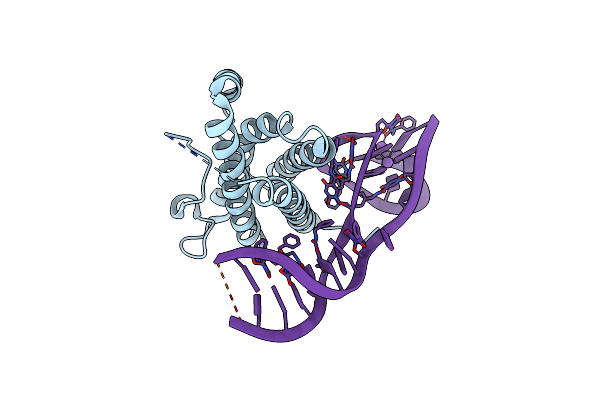 |
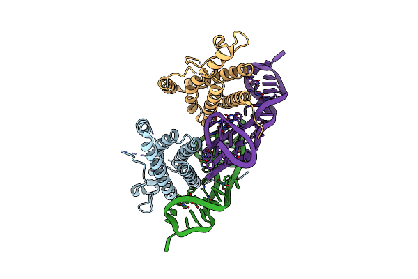
Crystal Structure Of Human Interleukin 6 In Complex With A Modified Nucleotide Aptamer (Somamer Sl1025)
Organism: Homo sapiens
Method: X-RAY DIFFRACTION Resolution:2.40 Å Release Date: 2014-01-22 Classification: CYTOKINE/DNA |
 |
Crystal Structure Of Human Interleukin 6 In Complex With A Modified Nucleotide Aptamer (Somamer Sl1025), Form 2
Organism: Homo sapiens
Method: X-RAY DIFFRACTION Resolution:2.55 Å Release Date: 2014-01-22 Classification: CYTOKINE/DNA |
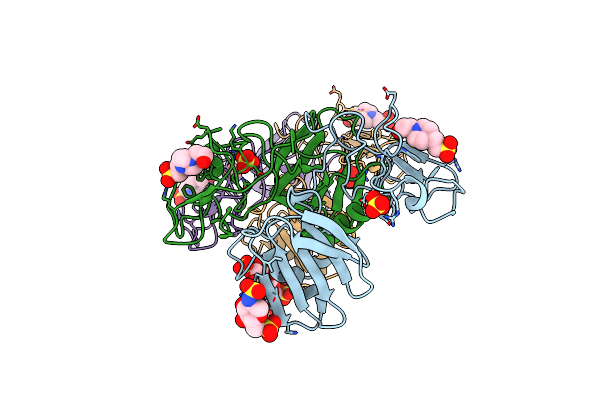 |
Crystal Structure Of 1K1 Mutant Of Hepatocyte Growth Factor/Scatter Factor Fragment Nk1 In Complex With Heparin
Organism: Homo sapiens
Method: X-RAY DIFFRACTION Resolution:2.81 Å Release Date: 2010-08-18 Classification: HORMONE Ligands: SO4, EPE, SGN |