
Search Count: 74
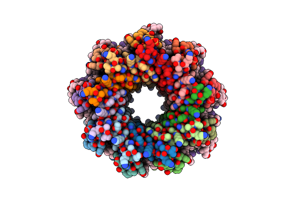 |
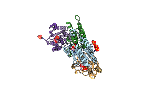
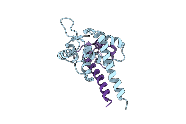
Organism: Rhodopseudomonas palustris atcc 17001
Method: ELECTRON MICROSCOPY Release Date: 2022-10-12 Classification: PHOTOSYNTHESIS Ligands: BCL, IRM |
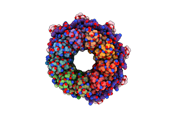 |
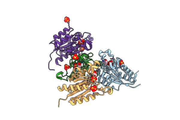
Organism: Rhodopseudomonas palustris
Method: ELECTRON MICROSCOPY Release Date: 2022-10-05 Classification: PHOTOSYNTHESIS Ligands: BCL, IRM |
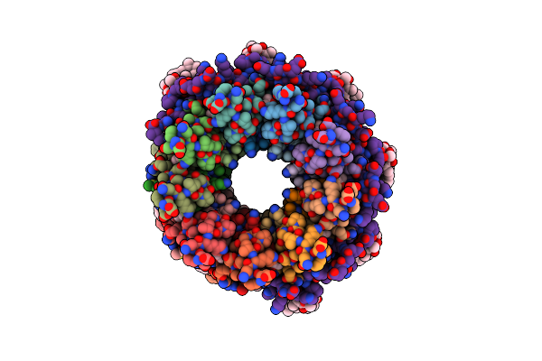 |
Organism: Rhodopseudomonas palustris
Method: ELECTRON MICROSCOPY Release Date: 2022-10-05 Classification: PHOTOSYNTHESIS Ligands: BCL, ZE0 |
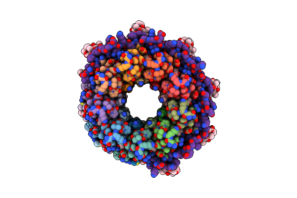 |
Organism: Rhodopseudomonas palustris
Method: ELECTRON MICROSCOPY Release Date: 2022-10-05 Classification: PHOTOSYNTHESIS Ligands: BCL, IRM |
 |
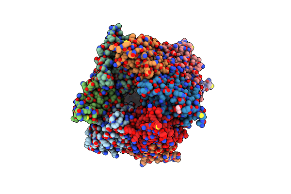
Crystal Structure Of Phycoerythrin From Cyanobacterium Nostoc Sp. Wr13 Contains Multiple Stacks Of Hexameric Assemblies Which Resemble The Rods Of Phycobilisome.
Organism: Nostoc sp. wr13
Method: X-RAY DIFFRACTION Resolution:2.13 Å Release Date: 2021-11-17 Classification: PHOTOSYNTHESIS Ligands: PEB, PG4, PGE, PEG, PO4, NO3, RWB, PE8, 1PE, P33, P6G, EDO, PE5 |
 |
The High Resolution Structure Of Allophycocyanin From Cyanobacterium Nostoc Sp. Wr13, The P21212 Crystal Form.
Organism: Nostoc sp. wr13
Method: X-RAY DIFFRACTION Resolution:1.42 Å Release Date: 2021-05-12 Classification: PHOTOSYNTHESIS Ligands: CYC, PG4, LYS, MPD, 1PE, PEG, EDO, P6G, GLU, GLY, ALA, MRD, PGE, DSN |
 |
The Structure Of Allophycocyanin From Cyanobacterium Nostoc Sp. Wr13, The C2221 Crystal Form.
Organism: Nostoc sp. wr13
Method: X-RAY DIFFRACTION Resolution:1.83 Å Release Date: 2021-05-12 Classification: PHOTOSYNTHESIS Ligands: PXQ, MPD, BCN, 1PE, PGE, PEG, EDO, PG4, MRD |
 |
The Crystal Structure Of Type Ii Dehydroquinase From Propionibacterium Acnes
Organism: Cutibacterium acnes
Method: X-RAY DIFFRACTION Resolution:1.70 Å Release Date: 2020-07-08 Classification: BIOSYNTHETIC PROTEIN Ligands: SO4, GOL |
 |
Organism: Zymomonas mobilis subsp. mobilis (strain atcc 31821 / zm4 / cp4)
Method: X-RAY DIFFRACTION Resolution:2.34 Å Release Date: 2020-07-08 Classification: BIOSYNTHETIC PROTEIN Ligands: FLC, TRS, SO4, GOL |
 |
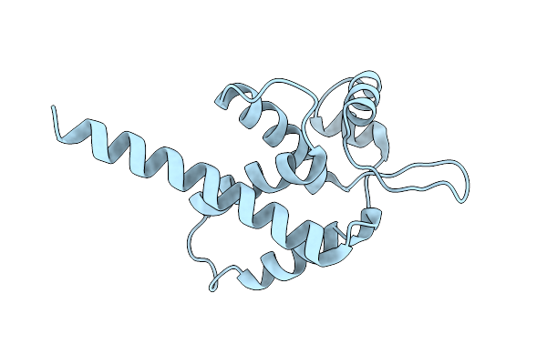
Organism: Phormidium rubidum a09dm
Method: X-RAY DIFFRACTION Resolution:1.17 Å Release Date: 2020-04-29 Classification: PHOTOSYNTHESIS Ligands: CYC, PEG, EDO, IMD, PGE |
 |
The Crystal Structure Of Type Ii Dehydroquinase From Butyrivibrio Crossotus Dsm 2876
Organism: Butyrivibrio crossotus dsm 2876
Method: X-RAY DIFFRACTION Resolution:1.70 Å Release Date: 2019-10-23 Classification: BIOSYNTHETIC PROTEIN Ligands: TLA, PO4, GOL |
 |
The Crystal Structure Of Type Ii Dehydroquinase From Butyrivibrio Crossotus Dsm 2876
Organism: Butyrivibrio crossotus dsm 2876
Method: X-RAY DIFFRACTION Resolution:1.05 Å Release Date: 2019-10-23 Classification: BIOSYNTHETIC PROTEIN Ligands: 3DS |
 |
The Crystal Structure Of Type Ii Dehydroquinase From Butyrivibrio Crossotus Dsm 2876
Organism: Butyrivibrio crossotus dsm 2876
Method: X-RAY DIFFRACTION Resolution:0.92 Å Release Date: 2019-10-23 Classification: BIOSYNTHETIC PROTEIN Ligands: ETE, SER, SO4, FLC, GOL |
 |
The Crystal Structure Of Type Ii Dehydroquinase From Acidithiobacillus Caldus Sm-1
Organism: Acidithiobacillus caldus (strain sm-1)
Method: X-RAY DIFFRACTION Resolution:1.95 Å Release Date: 2019-10-23 Classification: BIOSYNTHETIC PROTEIN Ligands: GOL, EPE |
 |
The Crystal Structure Of Type Ii Dehydroquinase From Psychromonas Ingrahamii 37 Crystal Form 1
Organism: Psychromonas ingrahamii (strain 37)
Method: X-RAY DIFFRACTION Resolution:1.46 Å Release Date: 2019-10-23 Classification: BIOSYNTHETIC PROTEIN Ligands: SO4, GOL |
 |
The Crystal Structure Of Type Ii Dehydroquinase From Psychromonas Ingrahamii 37, 40% Ethanol As Cryoprotectant
Organism: Psychromonas ingrahamii (strain 37)
Method: X-RAY DIFFRACTION Resolution:2.00 Å Release Date: 2019-10-23 Classification: BIOSYNTHETIC PROTEIN Ligands: EOH, SO4 |
 |
The Crystal Structure Of Type Ii Dehydroquinase From Psychromonas Ingrahamii 37, Crystal Form 2
Organism: Psychromonas ingrahamii (strain 37), Psychromonas ingrahamii 37
Method: X-RAY DIFFRACTION Resolution:1.60 Å Release Date: 2019-10-23 Classification: BIOSYNTHETIC PROTEIN Ligands: TLA, GOL, MPO |
 |
Hijacking The Hijackers: Escherichia Coli Pathogenicity Islands Redirect Helper Phage Packaging For Their Own Benefit.
Organism: Escherichia coli
Method: X-RAY DIFFRACTION Resolution:2.42 Å Release Date: 2019-07-31 Classification: DNA BINDING PROTEIN |
 |
Hijacking The Hijackers: Escherichia Coli Pathogenicity Islands Redirect Helper Phage Packaging For Their Own Benefit.
Organism: Escherichia coli, Escherichia virus lambda
Method: X-RAY DIFFRACTION Resolution:3.00 Å Release Date: 2019-07-31 Classification: DNA BINDING PROTEIN |
 |
C-Phycocyanin From Heterocyst Forming Filamentous Cyanobacterium Nostoc Sp. Wr13
Organism: Nostoc sp.
Method: X-RAY DIFFRACTION Resolution:1.51 Å Release Date: 2019-06-05 Classification: PHOTOSYNTHESIS Ligands: CYC, PG4, PEG, PO4, PGE, NO3, MPD |