
Search Count: 28
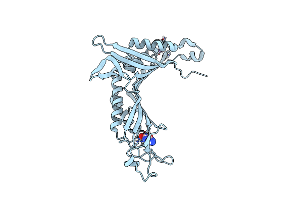 |
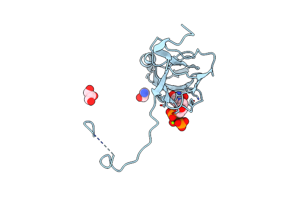
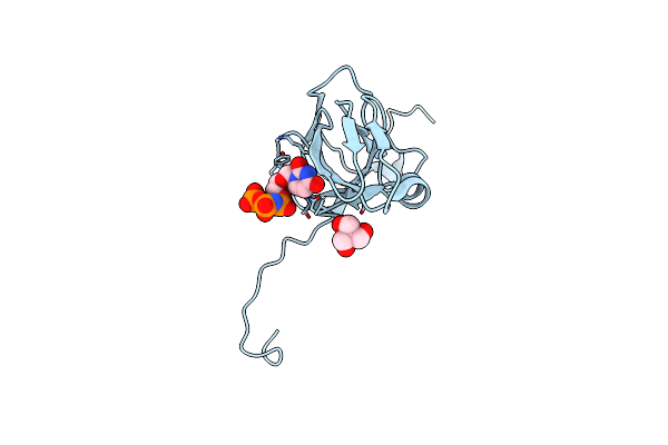
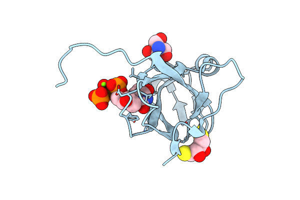
Joint Neutron/X-Ray Room Temperature Structure Of Perdeuterated Aspergillus Flavus Urate Oxidase In Complex With The 8-Azaxanthine Inhibitor And Catalytic Water Bound In The Peroxo Hole
Organism: Aspergillus flavus
Method: X-RAY DIFFRACTION, NEUTRON DIFFRACTION Resolution:1.33 Å, 2.10 Å, Release Date: 2020-12-09 Classification: OXIDOREDUCTASE Ligands: AZA, NA |
 |
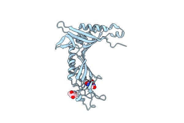
Organism: Simian immunodeficiency virus
Method: ELECTRON MICROSCOPY Release Date: 2020-02-05 Classification: RECOMBINATION Ligands: ZN |
 |
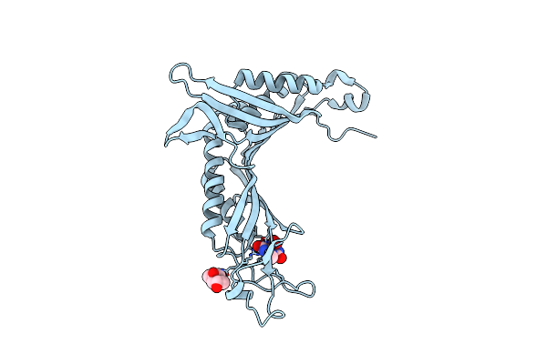
Organism: Simian immunodeficiency virus
Method: ELECTRON MICROSCOPY Release Date: 2020-02-05 Classification: RECOMBINATION Ligands: ZN, MG, KLQ, CL |
 |
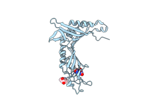
Organism: Simian immunodeficiency virus
Method: ELECTRON MICROSCOPY Release Date: 2020-02-05 Classification: RECOMBINATION Ligands: ZN, CL, DLU, MG |
 |
Organism: Simian immunodeficiency virus
Method: ELECTRON MICROSCOPY Release Date: 2020-02-05 Classification: RECOMBINATION Ligands: ZN, CL, KLQ, MG |
 |
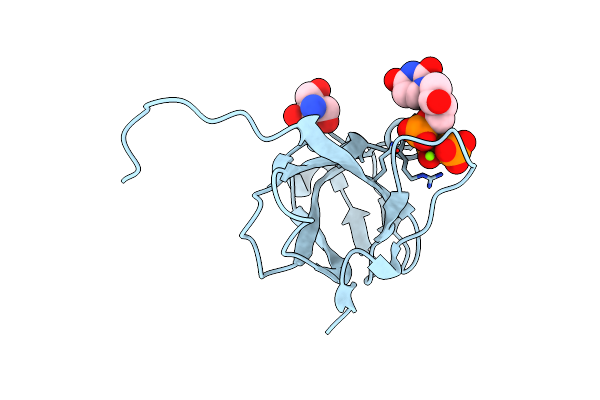
Organism: Mycobacterium tuberculosis
Method: X-RAY DIFFRACTION Resolution:1.30 Å Release Date: 2016-11-02 Classification: HYDROLASE Ligands: DUP, TRS, MG, GOL |
 |
Crystal Structure Of Mycobacterium Tuberculosis Dutpase R140K, H145W Mutant
Organism: Mycobacterium tuberculosis
Method: X-RAY DIFFRACTION Resolution:1.97 Å Release Date: 2016-11-02 Classification: HYDROLASE Ligands: TRS, DUP, MG |
 |
Crystal Structure Of Cofactor-Free Urate Oxidase In Complex With Its 5-Peroxoisourate Intermediate (X-Ray Dose, 106 Kgy)
Organism: Aspergillus flavus
Method: X-RAY DIFFRACTION Resolution:1.30 Å Release Date: 2014-11-05 Classification: OXIDOREDUCTASE Ligands: IUP, MPD, OXY, URC |
 |
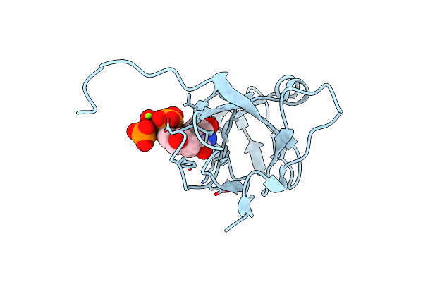
Crystal Structure Of Cofactor-Free Urate Oxidase Anaerobically Complexed With 9-Methyl Uric Acid
Organism: Aspergillus flavus
Method: X-RAY DIFFRACTION Resolution:1.50 Å Release Date: 2014-10-29 Classification: OXIDOREDUCTASE Ligands: MPD, MUA |
 |
Crystal Structure Of Cofactor-Free Urate Oxidase In Complex With The 5-Peroxo Derivative Of 9-Metyl Uric Acid (X-Ray Dose, 2.5 Kgy)
Organism: Aspergillus flavus
Method: X-RAY DIFFRACTION Resolution:1.32 Å Release Date: 2014-10-29 Classification: OXIDOREDUCTASE Ligands: XDS, MPD |
 |
Crystal Structure Of Cofactor-Free Urate Oxidase In Complex With The 5-Peroxo Derivative Of 9-Metyl Uric Acid (X-Ray Dose, 665 Kgy)
Organism: Aspergillus flavus
Method: X-RAY DIFFRACTION Resolution:1.34 Å Release Date: 2014-10-29 Classification: OXIDOREDUCTASE Ligands: XDS, MPD, OXY, MUA |
 |
Crystal Structure Of Cofactor-Free Urate Oxidase In Complex With The 5-Peroxo Derivative Of 9-Metyl Uric Acid (X-Ray Dose, 92 Kgy)
Organism: Aspergillus flavus
Method: X-RAY DIFFRACTION Resolution:1.28 Å Release Date: 2014-10-29 Classification: OXIDOREDUCTASE Ligands: XDS, MPD, OXY, MUA |
 |
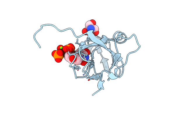
Crystal Structure Of Cofactor-Free Urate Oxidase Anaerobically Complexed With Uric Acid
Organism: Aspergillus flavus
Method: X-RAY DIFFRACTION Resolution:1.40 Å Release Date: 2014-10-29 Classification: OXIDOREDUCTASE Ligands: MPD, URC |
 |
Crystal Structure Of Cofactor-Free Urate Oxidase In Complex With Its 5-Peroxoisourate Intermediate (X-Ray Dose, 2.2 Kgy)
Organism: Aspergillus flavus
Method: X-RAY DIFFRACTION Resolution:1.30 Å Release Date: 2014-10-29 Classification: OXIDOREDUCTASE Ligands: IUP, MPD |
 |
Crystal Structure Of Cofactor-Free Urate Oxidase In Complex With Its 5-Peroxoisourate Intermediate (X-Ray Dose, 1.75 Mgy)
Organism: Aspergillus flavus
Method: X-RAY DIFFRACTION Resolution:1.35 Å Release Date: 2014-10-29 Classification: OXIDOREDUCTASE Ligands: IUP, MPD, OXY, URC |
 |
Organism: Mason-pfizer monkey virus
Method: X-RAY DIFFRACTION Resolution:1.65 Å Release Date: 2011-10-12 Classification: HYDROLASE Ligands: DUP, MG, TRS, DTT |
 |
Organism: Mason-pfizer monkey virus
Method: X-RAY DIFFRACTION Resolution:1.85 Å Release Date: 2011-10-12 Classification: HYDROLASE Ligands: MG, DUP, TRS |
 |
Crystal Structure Of M-Pmv Dutpase With A Mixed Population Of Substrate (Dupnpp) And Post-Inversion Product (Dump) In The Active Sites
Organism: Mason-pfizer monkey virus
Method: X-RAY DIFFRACTION Resolution:1.75 Å Release Date: 2011-10-12 Classification: HYDROLASE Ligands: MG, TRS, DUP, UMP |
 |
Crystal Structure Of M-Pmv Dutpase With A Mixed Population Of Substrate (Dupnpp) And Post-Inversion Product (Dump) In The Active Sites
Organism: Mason-pfizer monkey virus
Method: X-RAY DIFFRACTION Resolution:1.85 Å Release Date: 2011-10-12 Classification: HYDROLASE Ligands: UMP, DUP, MG |
 |
Crystal Structure Of M-Pmv Dutpase With A Mixed Population Of Substrate (Dupnpp) And Post-Inversion Product (Dump) In The Active Sites
Organism: Mason-pfizer monkey virus
Method: X-RAY DIFFRACTION Resolution:1.60 Å Release Date: 2011-10-12 Classification: HYDROLASE Ligands: UMP, MG, DUP, TRS |