
Search Count: 56
 |
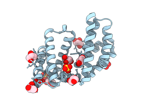
Organism: Synthetic construct
Method: X-RAY DIFFRACTION Release Date: 2025-12-10 Classification: DE NOVO PROTEIN Ligands: PGE, NA |
 |
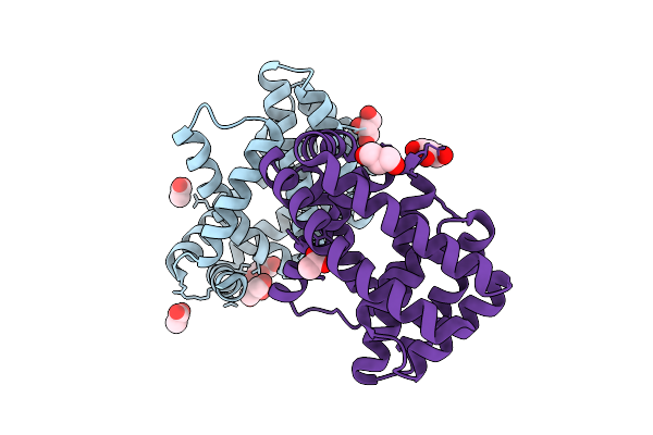
Organism: Synthetic construct
Method: X-RAY DIFFRACTION Release Date: 2025-12-10 Classification: DE NOVO PROTEIN Ligands: GOL, SO4 |
 |
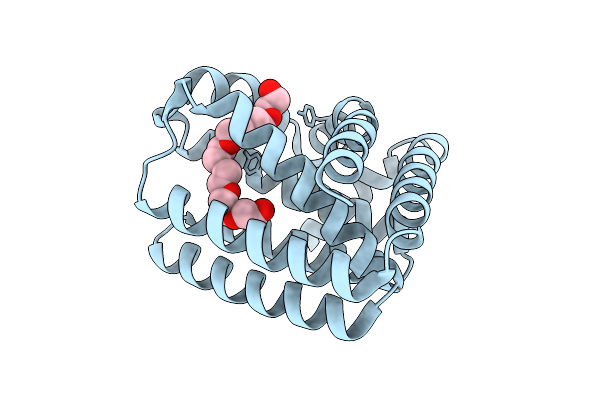
Organism: Synthetic construct
Method: X-RAY DIFFRACTION Release Date: 2025-08-13 Classification: DE NOVO PROTEIN Ligands: ACT, GOL, NA |
 |
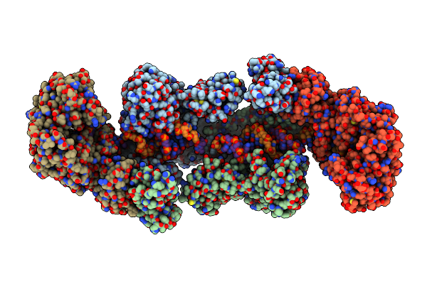
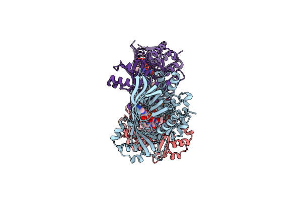
Organism: Vibrio cholerae, Vibrio cholerae o1
Method: ELECTRON MICROSCOPY Release Date: 2025-07-16 Classification: DNA BINDING PROTEIN/DNA Ligands: AGS, MG |
 |
Organism: Synthetic construct
Method: X-RAY DIFFRACTION Release Date: 2025-07-09 Classification: DE NOVO PROTEIN Ligands: 2PE |
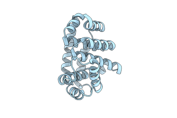 |
Organism: Synthetic construct
Method: X-RAY DIFFRACTION Release Date: 2025-07-09 Classification: DE NOVO PROTEIN |
 |
Organism: Synthetic construct
Method: X-RAY DIFFRACTION Release Date: 2025-07-09 Classification: DE NOVO PROTEIN Ligands: MG, AE4 |
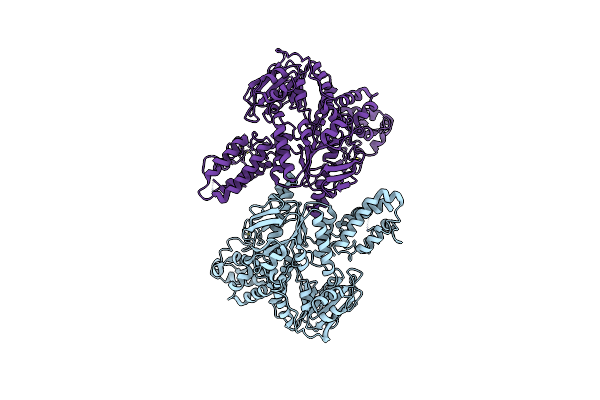 |
Organism: Mycobacterium tuberculosis
Method: ELECTRON MICROSCOPY Release Date: 2025-04-23 Classification: DNA BINDING PROTEIN Ligands: ZN |
 |
Organism: Mycobacterium tuberculosis, Escherichia coli
Method: ELECTRON MICROSCOPY Release Date: 2025-04-23 Classification: DNA BINDING PROTEIN Ligands: ZN, ADP |
 |
Organism: Mycobacterium tuberculosis, Escherichia coli
Method: ELECTRON MICROSCOPY Release Date: 2025-04-23 Classification: DNA BINDING PROTEIN Ligands: ZN |
 |
Organism: Mycobacterium tuberculosis, Escherichia coli
Method: ELECTRON MICROSCOPY Release Date: 2025-04-23 Classification: DNA BINDING PROTEIN Ligands: ZN, ADP |
 |
Organism: Neobacillus niacini
Method: X-RAY DIFFRACTION Resolution:3.14 Å Release Date: 2024-09-18 Classification: OXIDOREDUCTASE Ligands: FAD, DR9 |
 |
Organism: Neobacillus niacini
Method: ELECTRON MICROSCOPY Release Date: 2024-09-18 Classification: CYTOSOLIC PROTEIN Ligands: FAD, DR9 |
 |
Organism: Neobacillus niacini
Method: ELECTRON MICROSCOPY Release Date: 2024-09-18 Classification: CYTOSOLIC PROTEIN Ligands: FAD, WTQ, DR9 |
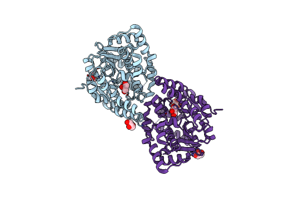 |
Structure Of A Cytosolic Sulfotransferase Of Anopheles Gambiae (Agap001425).
Organism: Anopheles gambiae
Method: X-RAY DIFFRACTION Resolution:2.00 Å Release Date: 2022-08-10 Classification: TRANSFERASE Ligands: GOL, CL |
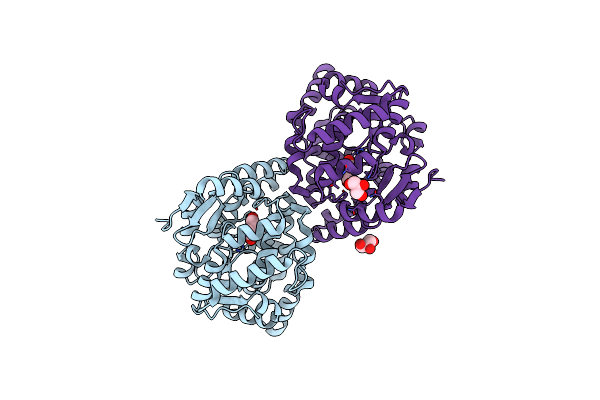 |
Structure Of A Cytosolic Sulfotransferase Of Anopheles Gambiae (Agap001425) In Complex With Vanillin
Organism: Anopheles gambiae
Method: X-RAY DIFFRACTION Resolution:2.10 Å Release Date: 2022-08-10 Classification: TRANSFERASE Ligands: V55, CL, GOL |
 |
Structure Of A Cytosolic Sulfotransferase Of Anopheles Gambiae (Agap001425) In Complex With 3'-Phosphoadenosine 5-Phosphate And Vanillin.
Organism: Anopheles gambiae
Method: X-RAY DIFFRACTION Resolution:2.50 Å Release Date: 2022-08-10 Classification: TRANSFERASE Ligands: V55, A3P, GOL |
 |
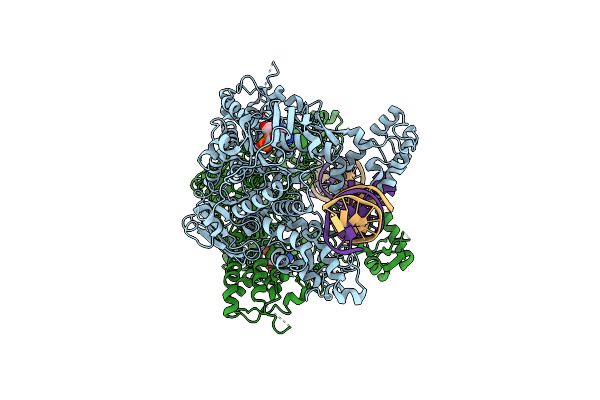
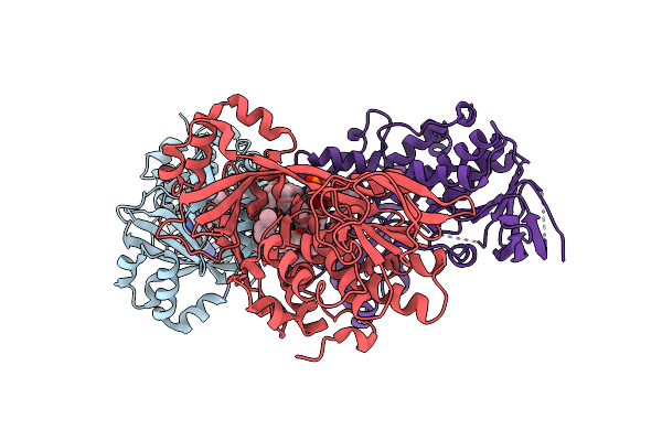
Crystal Structure Of Haemophilus Influenzae 3-Isopropylmalate Dehydrogenase In Complex With O-Isobutenyl Oxalylhydroxamate.
Organism: Haemophilus influenzae (strain atcc 51907 / dsm 11121 / kw20 / rd)
Method: X-RAY DIFFRACTION Resolution:2.09 Å Release Date: 2020-02-26 Classification: OXIDOREDUCTASE Ligands: NAD, MG, O45 |
 |
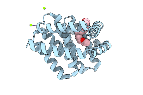
The Structure Of Ligand Binding Domain Of Lasr In Complex With Tp-1 Homolog, Compound 10
Organism: Pseudomonas aeruginosa (strain atcc 15692 / dsm 22644 / cip 104116 / jcm 14847 / lmg 12228 / 1c / prs 101 / pao1)
Method: X-RAY DIFFRACTION Resolution:1.90 Å Release Date: 2018-08-08 Classification: signaling protein/agonist Ligands: FXD |
 |
The Structure Of Ligand Binding Domain Of Lasr In Complex With Tp-1 Homolog, Compound 11
Organism: Pseudomonas aeruginosa (strain atcc 15692 / dsm 22644 / cip 104116 / jcm 14847 / lmg 12228 / 1c / prs 101 / pao1)
Method: X-RAY DIFFRACTION Resolution:1.70 Å Release Date: 2018-08-08 Classification: signaling protein/agonist Ligands: FXS |