
Search Count: 19
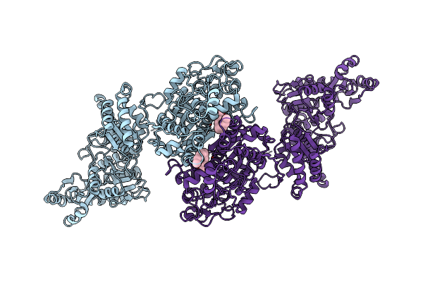 |
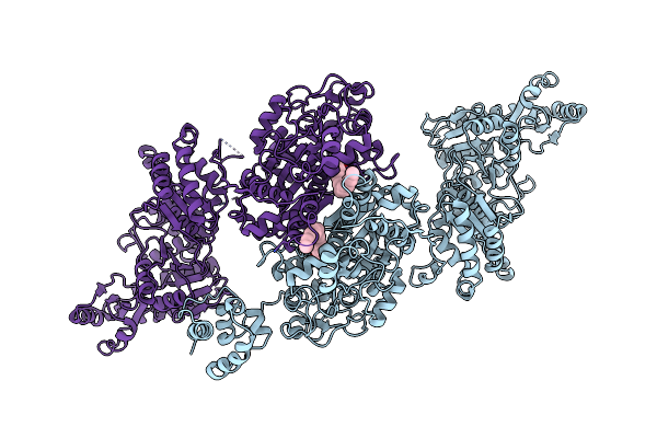
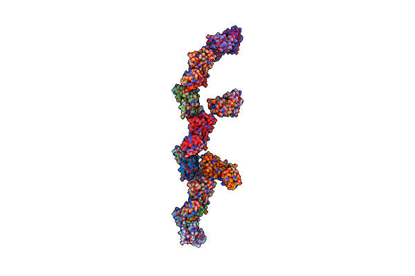
Organism: Mycolicibacterium smegmatis mc2 155
Method: ELECTRON MICROSCOPY Release Date: 2023-02-15 Classification: BIOSYNTHETIC PROTEIN Ligands: UNL |
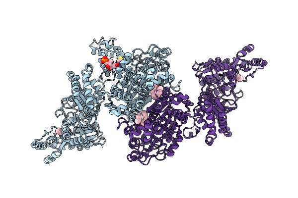 |
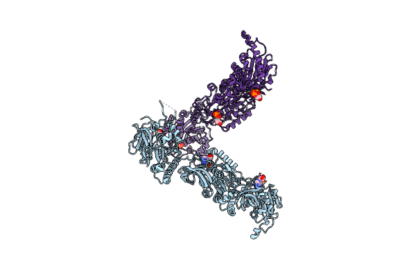
Organism: Mycolicibacterium smegmatis mc2 155
Method: ELECTRON MICROSCOPY Release Date: 2023-02-15 Classification: BIOSYNTHETIC PROTEIN Ligands: UNL, PNS |
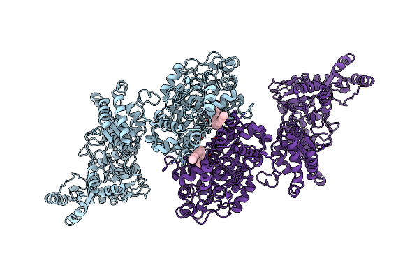 |
Organism: Mycolicibacterium smegmatis mc2 155
Method: ELECTRON MICROSCOPY Release Date: 2023-02-15 Classification: BIOSYNTHETIC PROTEIN Ligands: UNL |
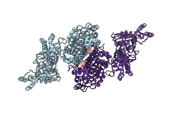 |
Organism: Mycolicibacterium smegmatis mc2 155
Method: ELECTRON MICROSCOPY Release Date: 2023-02-15 Classification: BIOSYNTHETIC PROTEIN Ligands: UNL |
 |
Organism: Mycolicibacterium smegmatis mc2 155
Method: ELECTRON MICROSCOPY Release Date: 2023-02-15 Classification: BIOSYNTHETIC PROTEIN Ligands: UNL |
 |
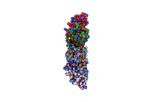
Sars-Cov-2 Wuhan-Hu-1-Spike-Rbd Bound To Linker Variant Of Affinity Matured Ace2 Mimetic Cvd432
Organism: Severe acute respiratory syndrome coronavirus 2, Homo sapiens
Method: ELECTRON MICROSCOPY Release Date: 2022-08-31 Classification: VIRAL PROTEIN/ANTIVIRAL PROTEIN Ligands: NAG |
 |
Sars-Cov-2 Wuhan-Hu-1-Spike-Rbd Bound To Computationally Engineered Ace2 Mimetic Cvd293
Organism: Severe acute respiratory syndrome coronavirus 2, Homo sapiens
Method: ELECTRON MICROSCOPY Release Date: 2022-08-31 Classification: VIRAL PROTEIN/ANTIVIRAL PROTEIN Ligands: NAG |
 |
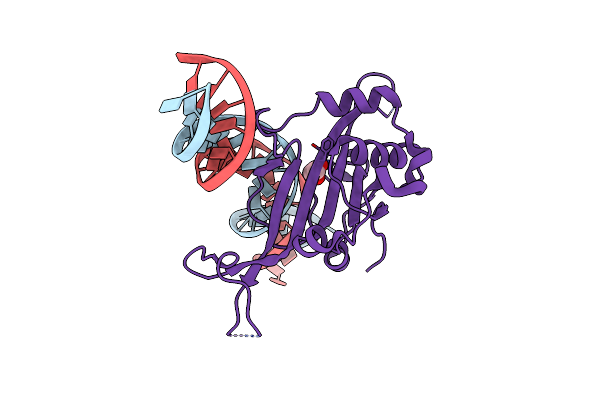
Organism: Homo sapiens, Severe acute respiratory syndrome coronavirus 2
Method: ELECTRON MICROSCOPY Release Date: 2020-10-21 Classification: VIRAL PROTEIN |
 |
Organism: Mycobacterium smegmatis (strain atcc 700084 / mc(2)155)
Method: ELECTRON MICROSCOPY Release Date: 2019-10-23 Classification: TRANSPORT PROTEIN |
 |
The Structure Of The C-Terminus Of Virulence Protein Ince From Chlamydia Trachomatis Bound To Mus Musculus Snx5-Px Domain
Organism: Mus musculus, Chlamydia trachomatis d/uw-3/cx
Method: X-RAY DIFFRACTION Resolution:2.31 Å Release Date: 2017-08-30 Classification: PROTEIN TRANSPORT |
 |
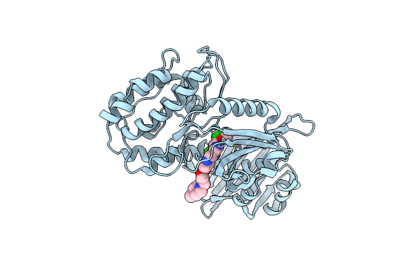
Thermomonospora Curvata Eccc (Atpases 2 And 3) In Complex With A Signal Sequence Peptide
Organism: Thermomonospora curvata
Method: X-RAY DIFFRACTION Resolution:3.24 Å Release Date: 2015-02-11 Classification: PROTEIN BINDING/PROTEIN BINDING Ligands: ATP, MG |
 |
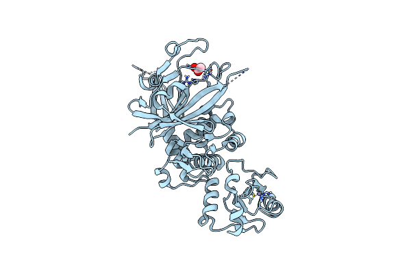
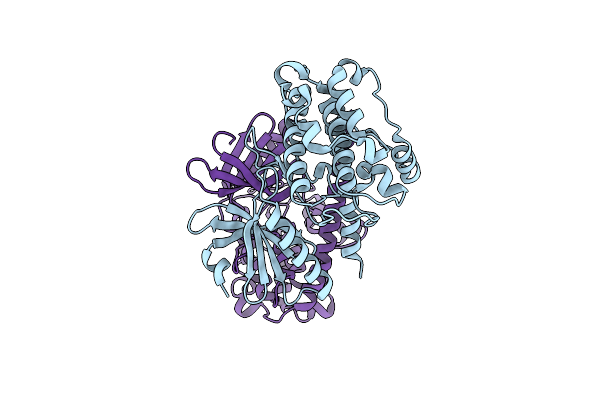
Cytoplasmic Domain Of The Thermomonospora Curvata Type Vii Secretion Atpase Eccc
Organism: Thermomonospora curvata
Method: X-RAY DIFFRACTION Resolution:2.90 Å Release Date: 2015-02-11 Classification: CELL CYCLE Ligands: SO4, MG, ATP |
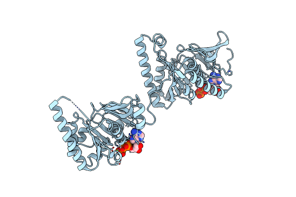 |
Organism: Geobacillus thermodenitrificans
Method: X-RAY DIFFRACTION Resolution:2.45 Å Release Date: 2015-02-04 Classification: CELL CYCLE Ligands: MG, ATP |
 |
Organism: Saccharomyces cerevisiae
Method: X-RAY DIFFRACTION Resolution:2.61 Å Release Date: 2014-06-25 Classification: TRANSCRIPTION/DNA Ligands: EDO |
 |
Organism: Saccharomyces cerevisiae
Method: X-RAY DIFFRACTION Resolution:1.87 Å Release Date: 2014-04-16 Classification: SPLICING Ligands: ZN, ACT |
 |
Organism: Homo sapiens
Method: X-RAY DIFFRACTION Resolution:3.55 Å Release Date: 2011-08-31 Classification: TRANSFERASE/TRANSFERASE INHIBITOR Ligands: DB8 |
 |
Crystal Structure Of A Key Regulator Of Virulence In Mycobacterium Tuberculosis
Organism: Mycobacterium tuberculosis
Method: X-RAY DIFFRACTION Resolution:2.50 Å Release Date: 2011-08-03 Classification: TRANSCRIPTION |
 |
Organism: Caenorhabditis elegans
Method: X-RAY DIFFRACTION Resolution:2.64 Å Release Date: 2006-02-07 Classification: TRANSFERASE |
 |
Crystal Structure Of The Auto-Inhibited Kinase Domain Of Calcium/Calmodulin Activated Kinase Ii
Organism: Caenorhabditis elegans
Method: X-RAY DIFFRACTION Resolution:1.80 Å Release Date: 2005-12-13 Classification: TRANSFERASE |