
Search Count: 12
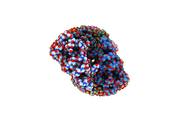 |
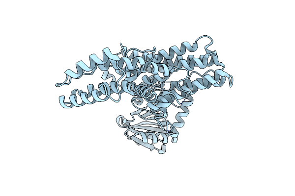
Subtomogram Averaging Structure Of Cofilactin Filament Inside Microtubule Lumen Of Drosophila S2 Cell Protrusion.
Organism: Drosophila
Method: ELECTRON MICROSCOPY Release Date: 2023-05-10 Classification: CONTRACTILE PROTEIN |
 |
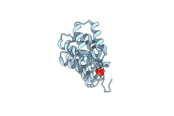
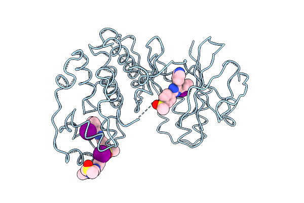
Organism: Homo sapiens
Method: X-RAY DIFFRACTION Resolution:2.20 Å Release Date: 2017-03-15 Classification: OXIDOREDUCTASE, LYASE/INHIBITOR Ligands: HEM, 6D7, 7D6 |
 |
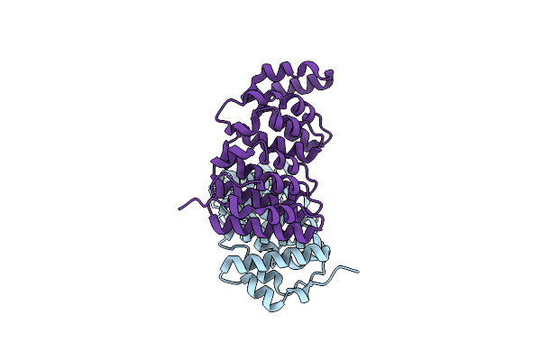
Organism: Homo sapiens
Method: X-RAY DIFFRACTION Resolution:3.10 Å Release Date: 2017-03-15 Classification: OXIDOREDUCTASE, LYASE/INHIBITOR Ligands: HEM, 6D8 |
 |
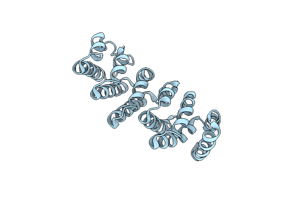
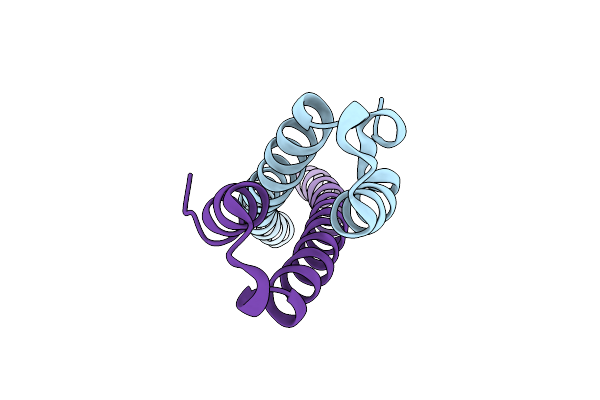
The Xmap215 Family Drives Microtubule Polymerization Using A Structurally Diverse Tog Array
Organism: Drosophila melanogaster
Method: X-RAY DIFFRACTION Resolution:1.65 Å Release Date: 2014-07-09 Classification: PROTEIN BINDING Ligands: SO4 |
 |
The Xmap215 Family Drives Microtubule Polymerization Using A Structurally Diverse Tog Array
Organism: Homo sapiens
Method: X-RAY DIFFRACTION Resolution:1.90 Å Release Date: 2014-07-09 Classification: PROTEIN BINDING |
 |
The Xmap215 Family Drives Microtubule Polymerization Using A Structurally Diverse Tog Array
Organism: Homo sapiens
Method: X-RAY DIFFRACTION Resolution:2.50 Å Release Date: 2014-07-09 Classification: PROTEIN BINDING |
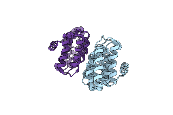 |
A Cryptic Tog Domain With A Distinct Architecture Underlies Clasp-Dependent Bipolar Spindle Formation
Organism: Homo sapiens
Method: X-RAY DIFFRACTION Resolution:2.01 Å Release Date: 2013-06-12 Classification: STRUCTURAL PROTEIN |
 |
Organism: Entamoeba histolytica
Method: X-RAY DIFFRACTION Resolution:2.30 Å Release Date: 2013-01-09 Classification: SIGNALING PROTEIN |
 |
Organism: Homo sapiens
Method: X-RAY DIFFRACTION Resolution:1.80 Å Release Date: 2005-03-08 Classification: STRUCTURAL PROTEIN |
 |
Organism: Homo sapiens
Method: X-RAY DIFFRACTION Resolution:2.00 Å Release Date: 2005-03-08 Classification: STRUCTURAL PROTEIN |
 |
Organism: Homo sapiens
Method: X-RAY DIFFRACTION Resolution:2.00 Å Release Date: 1998-05-06 Classification: SERINE/THREONINE-PROTEIN KINASE Ligands: D13 |
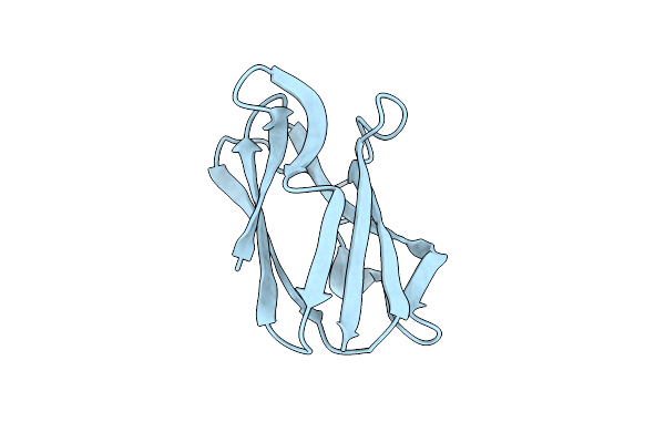 |
The Crystal Structure Of Poplar Apoplastocyanin At 1.8-Angstroms Resolution. The Geometry Of The Copper-Binding Site Is Created By The Polypeptide
Organism: Populus nigra
Method: X-RAY DIFFRACTION Resolution:1.80 Å Release Date: 1984-02-02 Classification: ELECTRON TRANSPORT PROTEIN(CUPROPROTEIN) |