
Search Count: 33
All
Selected
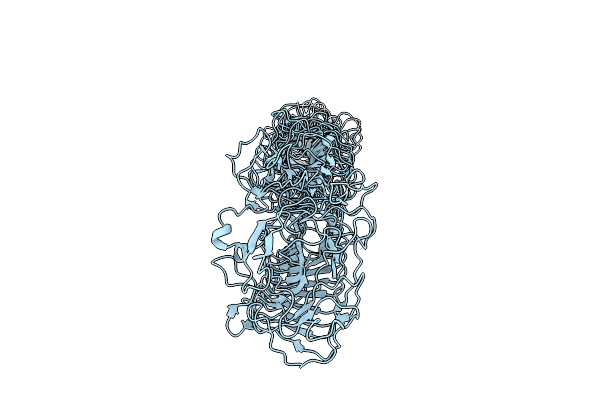 |
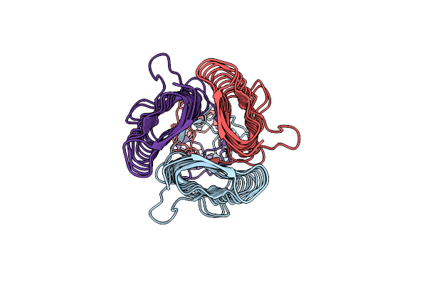
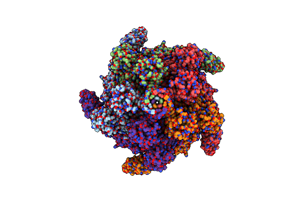
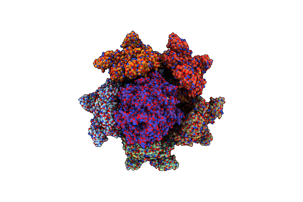
Membrane-Distal Part Of Extracellular Domain Of The Fap2 Autotransporter Adhesin From Fusobacterium Nucleatum Atcc23726
Organism: Fusobacterium nucleatum
Method: ELECTRON MICROSCOPY Resolution:4.40 Å Release Date: 2025-07-30 Classification: CELL ADHESION |
 |
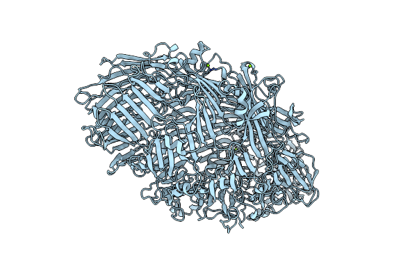
Organism: Homo sapiens
Method: ELECTRON MICROSCOPY Release Date: 2025-06-11 Classification: TRANSFERASE |
 |
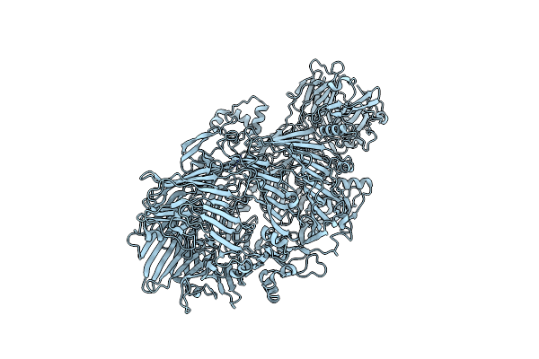
Organism: Homo sapiens
Method: ELECTRON MICROSCOPY Resolution:3.30 Å Release Date: 2025-06-11 Classification: TRANSFERASE |
 |
Organism: Homo sapiens
Method: ELECTRON MICROSCOPY Resolution:3.80 Å Release Date: 2025-06-11 Classification: TRANSFERASE |
 |
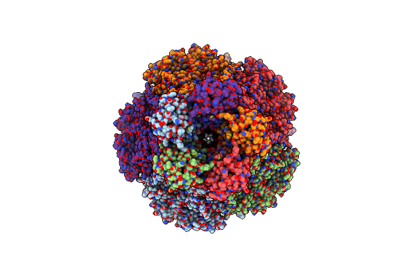
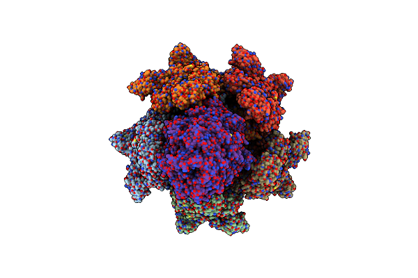
Organism: Fusobacterium nucleatum
Method: ELECTRON MICROSCOPY Release Date: 2025-04-02 Classification: CELL ADHESION |
 |
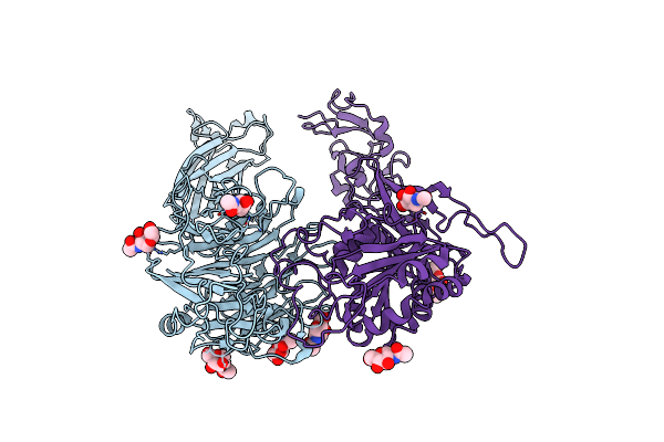
Organism: Fusobacterium nucleatum, Homo sapiens
Method: ELECTRON MICROSCOPY Release Date: 2025-04-02 Classification: CELL ADHESION Ligands: NAG |
 |
Organism: Homo sapiens
Method: ELECTRON MICROSCOPY Release Date: 2025-04-02 Classification: CELL ADHESION Ligands: NAG |
 |
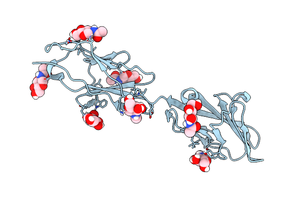
Organism: Rattus norvegicus
Method: ELECTRON MICROSCOPY Release Date: 2024-11-13 Classification: CELL ADHESION Ligands: NAG |
 |
Organism: Photorhabdus luminescens
Method: ELECTRON MICROSCOPY Release Date: 2024-03-13 Classification: TOXIN |
 |
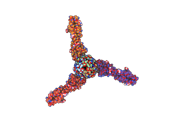
Photorhabdus Luminescens Tcda1 Prepore-To-Pore Intermediate, K567W K2008W Mutant
Organism: Photorhabdus luminescens
Method: ELECTRON MICROSCOPY Release Date: 2024-03-13 Classification: TOXIN |
 |
Photorhabdus Luminescens Tcda1 Prepore-To-Pore Intermediate, C16S, C20S, C870S, T1279C Mutant
Organism: Photorhabdus luminescens
Method: ELECTRON MICROSCOPY Release Date: 2024-03-13 Classification: TOXIN |
 |
Organism: Photorhabdus luminescens, Oryctolagus cuniculus
Method: ELECTRON MICROSCOPY Release Date: 2022-06-29 Classification: TOXIN Ligands: ADP, MG, APR, NCA |
 |
Organism: Oryctolagus cuniculus
Method: ELECTRON MICROSCOPY Release Date: 2022-06-29 Classification: CYTOSOLIC PROTEIN Ligands: ADP, MG, APR |
 |
Structure Of The Adp-Ribosyltransferase Tccc3Hvr From Photorhabdus Luminescens
Organism: Photorhabdus luminescens
Method: SOLUTION NMR Release Date: 2022-06-29 Classification: TOXIN |
 |
Organism: Morganella morganii
Method: ELECTRON MICROSCOPY Release Date: 2020-07-08 Classification: TOXIN |
 |
Organism: Xenorhabdus nematophila
Method: ELECTRON MICROSCOPY Release Date: 2020-07-08 Classification: TOXIN |
 |
Organism: Photorhabdus luminescens, Homo sapiens
Method: X-RAY DIFFRACTION Resolution:2.00 Å Release Date: 2019-12-04 Classification: TOXIN Ligands: MG |
 |
Organism: Photorhabdus luminescens, Tobacco etch virus
Method: X-RAY DIFFRACTION Resolution:3.70 Å Release Date: 2019-12-04 Classification: TOXIN |
 |
Structure Of Photorhabdus Luminescens Tc Holotoxin Pore, Mutation Tccc3-D651A
Organism: Photorhabdus luminescens
Method: ELECTRON MICROSCOPY Release Date: 2019-11-06 Classification: TOXIN |
 |
Organism: Photorhabdus luminescens
Method: ELECTRON MICROSCOPY Release Date: 2019-11-06 Classification: TOXIN |