
Search Count: 53
 |
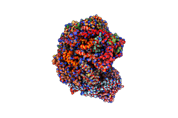
Organism: Influenza a virus, Aequorea victoria
Method: ELECTRON MICROSCOPY Release Date: 2025-08-13 Classification: VIRAL PROTEIN Ligands: NAG |
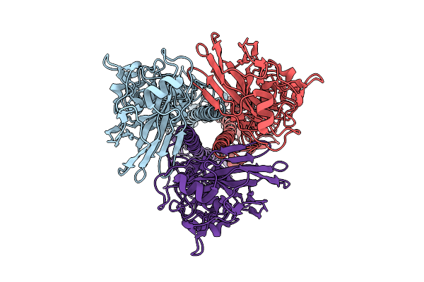 |
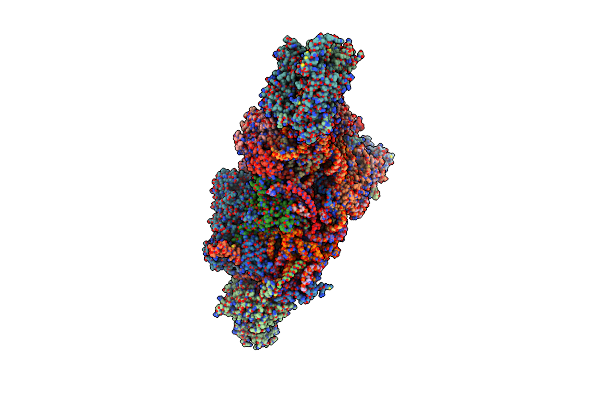
Organism: Influenza a virus
Method: ELECTRON MICROSCOPY Release Date: 2025-08-13 Classification: VIRAL PROTEIN |
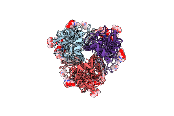 |
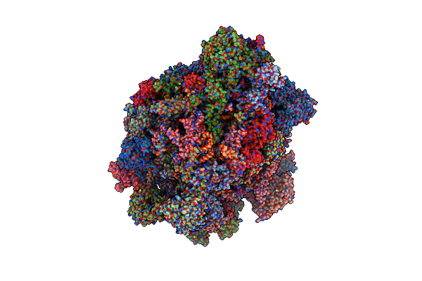
Organism: Influenza a virus
Method: ELECTRON MICROSCOPY Release Date: 2025-08-13 Classification: VIRAL PROTEIN Ligands: NAG |
 |
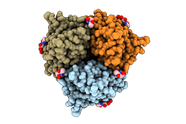
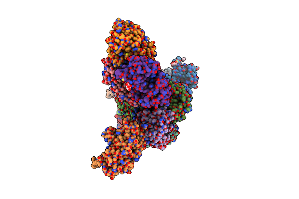
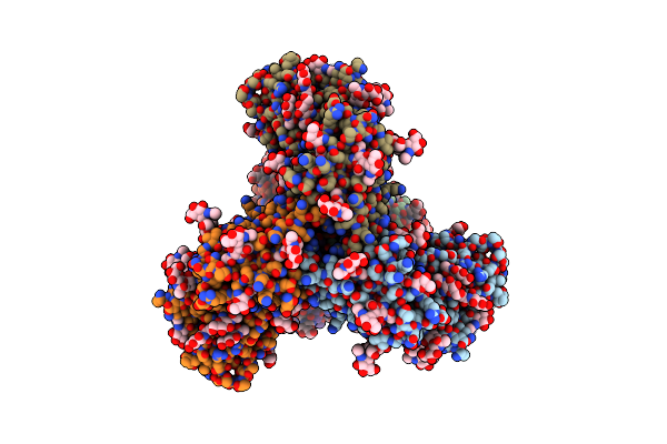
Organism: Influenza a virus (strain a/hong kong/1/1968 h3n2), Aequorea victoria
Method: ELECTRON MICROSCOPY Release Date: 2025-08-06 Classification: VIRAL PROTEIN Ligands: NAG |
 |
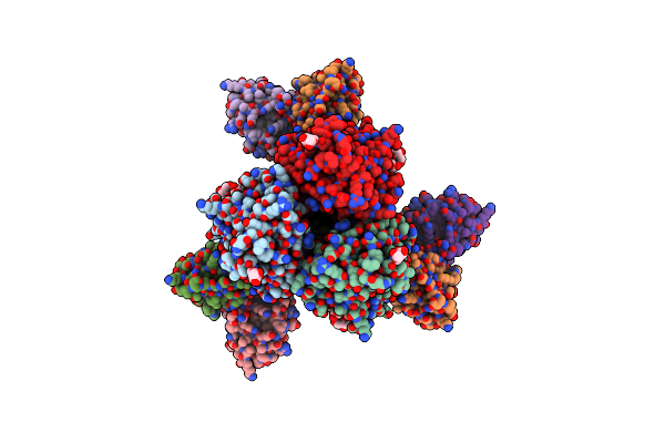
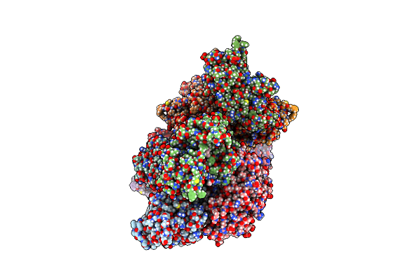
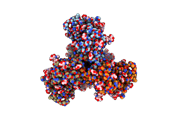
Organism: Influenza a virus (strain a/hong kong/1/1968 h3n2), Aequorea victoria
Method: ELECTRON MICROSCOPY Release Date: 2025-08-06 Classification: VIRAL PROTEIN Ligands: NAG |
 |
Organism: Human coronavirus oc43
Method: ELECTRON MICROSCOPY Release Date: 2025-04-23 Classification: VIRAL PROTEIN Ligands: NAG |
 |
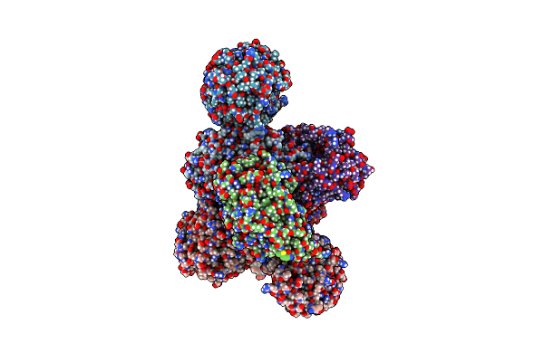
Hemagglutinin H1 New Caledonia 1999 In Complex With Monoclonal Antibody Fab 43_S0008
Organism: Influenza a virus, Mus sp.
Method: ELECTRON MICROSCOPY Release Date: 2024-12-11 Classification: VIRAL PROTEIN/IMMUNE SYSTEM Ligands: NAG |
 |
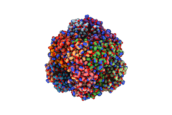
Ssu(Head) Structure Derived From The Ssu Sample Of The Mitoribosome From T. Gondii.
Organism: Toxoplasma gondii
Method: ELECTRON MICROSCOPY Release Date: 2024-12-11 Classification: RIBOSOME |
 |
Ssu(Body) Structure Derived From The Ssu Sample Of The Mitoribosome From T. Gondii.
Organism: Toxoplasma gondii
Method: ELECTRON MICROSCOPY Release Date: 2024-12-11 Classification: RIBOSOME |
 |
Lsu Structure Derived From The Lsu Sample Of The Mitoribosome From T. Gondii.
Organism: Toxoplasma gondii
Method: ELECTRON MICROSCOPY Release Date: 2024-12-11 Classification: RIBOSOME |
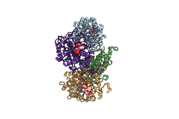 |
Succinate Bound Crystal Structure Of Thermus Scotoductus Sa-01 Ene-Reductase
Organism: Thermus scotoductus sa-01
Method: X-RAY DIFFRACTION Resolution:2.14 Å Release Date: 2024-07-03 Classification: OXIDOREDUCTASE Ligands: SIN, FMN |
 |
Rhodococcus Ruber Alcohol Dehydrogenase Nadh Biomimetic Complex - Compound 4B
Organism: Rhodococcus ruber
Method: X-RAY DIFFRACTION Resolution:2.99 Å Release Date: 2024-07-03 Classification: OXIDOREDUCTASE Ligands: ZN, W46, CIT, IPA |
 |
Rhodococcus Ruber Alcohol Dehydrogenase Nadh Biomimetic Complex - Compound 1A
Organism: Rhodococcus ruber
Method: X-RAY DIFFRACTION Resolution:2.20 Å Release Date: 2024-07-03 Classification: OXIDOREDUCTASE Ligands: IPA, ZN, NA, W3O, CIT |
 |
Organism: Thermus scotoductus sa-01
Method: X-RAY DIFFRACTION Resolution:2.76 Å Release Date: 2024-07-03 Classification: FLAVOPROTEIN Ligands: FMN, W3X, IPA, NA, CL |
 |
Low Resolution Structure Of Inactive Conformation Of The Ktr Cation Channel In Presence Of Atp And C-Di-Amp
Organism: Bacillus subtilis subsp. subtilis str. 168
Method: X-RAY DIFFRACTION Resolution:5.77 Å Release Date: 2024-05-15 Classification: MEMBRANE PROTEIN |
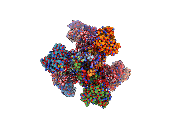 |
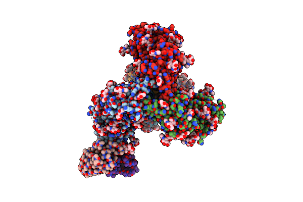
Crystal Structure Of The Bg505 Triple Tandem Trimer Gp140 Hiv-1 Env In Complex With Pgt124 And 35O22
Organism: Human immunodeficiency virus 1, Homo sapiens
Method: X-RAY DIFFRACTION Resolution:5.75 Å Release Date: 2024-04-17 Classification: VIRAL PROTEIN/IMMUNE SYSTEM Ligands: NAG |
 |
Bg505 Sosip Reconstructed From A Designed Nanoparticle, Bg505 Sosip-T33-31 (Component A)
Organism: Human immunodeficiency virus 1
Method: ELECTRON MICROSCOPY Release Date: 2021-08-04 Classification: VIRAL PROTEIN Ligands: NAG |
 |
Bg505 Sosip Reconstructed From A Designed Nanoparticle, Bg505 Sosip-T33-31 (Component B)
Organism: Human immunodeficiency virus 1
Method: ELECTRON MICROSCOPY Release Date: 2021-08-04 Classification: VIRAL PROTEIN Ligands: NAG |
 |
Organism: Human immunodeficiency virus 1
Method: ELECTRON MICROSCOPY Release Date: 2021-08-04 Classification: VIRAL PROTEIN |
 |
Bg505 Sosip Md39 In Complex With The Polyclonal Fab Pabc-1 From Animal Rh.32034 (Wk26 Time Point)
Organism: Human immunodeficiency virus 1, Macaca mulatta
Method: ELECTRON MICROSCOPY Release Date: 2021-08-04 Classification: VIRAL PROTEIN/Immune System Ligands: NAG |