
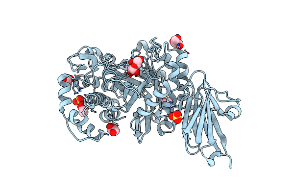
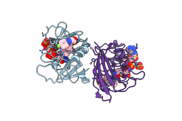
Search Count: 23
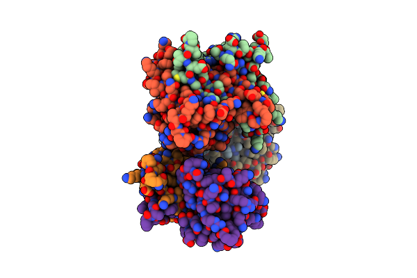 |
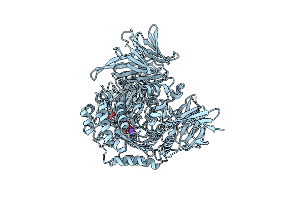
Organism: Severe acute respiratory syndrome coronavirus 2
Method: X-RAY DIFFRACTION Resolution:1.85 Å Release Date: 2022-06-22 Classification: RNA BINDING PROTEIN, Viral protein Ligands: GOL, ACT |
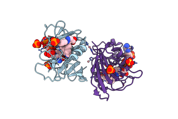
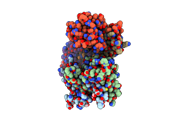 |
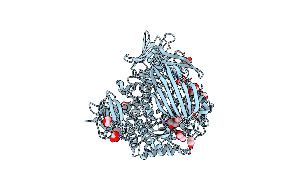
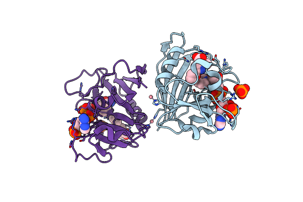
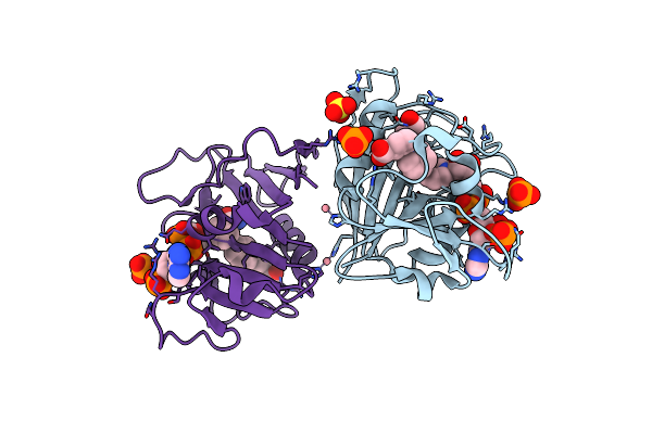
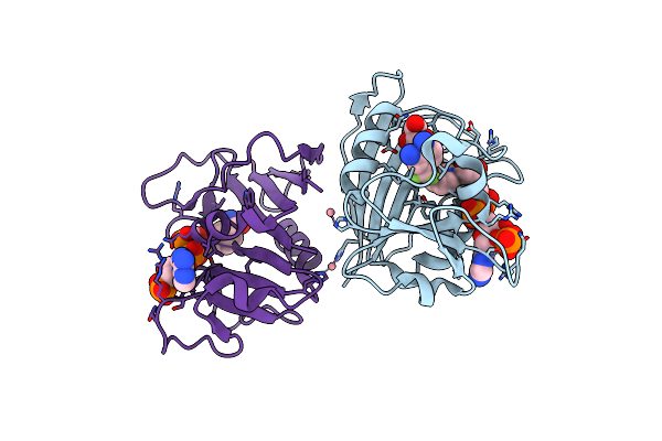
Crystal Structure Of Sars-Cov-2 Nucleocapsid Protein C-Terminal Domain Complexed With Chicoric Acid
Organism: Severe acute respiratory syndrome coronavirus 2
Method: X-RAY DIFFRACTION Resolution:1.73 Å Release Date: 2022-06-22 Classification: RNA BINDING PROTEIN/Viral Protein Ligands: GKP, PEG, GOL, NA, CL |
 |
Crystal Structure Of Xac1771, A Novel Carbohydrate Acetylesterase From Xanthomonas Citri
Organism: Xanthomonas axonopodis pv. citri (strain 306)
Method: X-RAY DIFFRACTION Resolution:1.90 Å Release Date: 2021-05-26 Classification: HYDROLASE Ligands: ZN |
 |
Crystal Structure Of Xac1772, A Gh35 Xyloglucan-Active Beta-Galactosidase From Xanthomonas Citri
Organism: Xanthomonas axonopodis pv. citri (strain 306)
Method: X-RAY DIFFRACTION Resolution:1.80 Å Release Date: 2021-05-26 Classification: HYDROLASE Ligands: SO4 |
 |
Crystal Structure Of The Gh35 Beta-Galactosidase (Xac1772) From Xanthomonas Citri In Complex With Galactose
Organism: Xanthomonas axonopodis pv. citri (strain 306)
Method: X-RAY DIFFRACTION Resolution:1.75 Å Release Date: 2021-05-26 Classification: HYDROLASE Ligands: SO4, GLA, GOL |
 |
Crystal Structure Of The Gh31 Alpha-Xylosidase (Xac1773) From Xanthomonas Citri
Organism: Xanthomonas axonopodis pv. citri (strain 306)
Method: X-RAY DIFFRACTION Resolution:1.56 Å Release Date: 2021-05-26 Classification: HYDROLASE Ligands: GOL, K |
 |
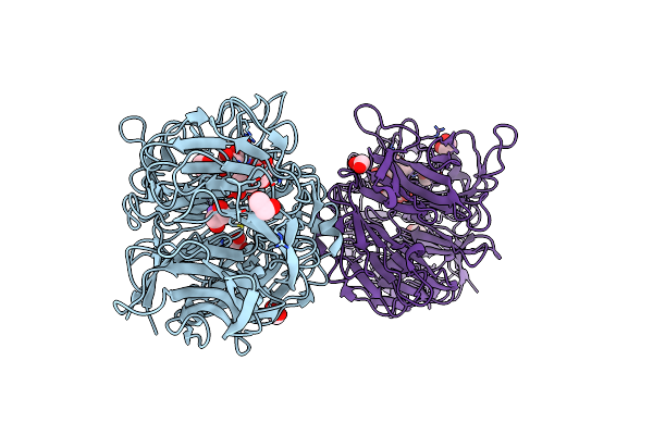
Crystal Structure Of The Gh95 Alpha-L-1,2-Fucosidase (Xac1774) From Xanthomonas Citri
Organism: Xanthomonas axonopodis pv. citri (strain 306)
Method: X-RAY DIFFRACTION Resolution:2.05 Å Release Date: 2021-05-26 Classification: HYDROLASE Ligands: GOL, CA |
 |
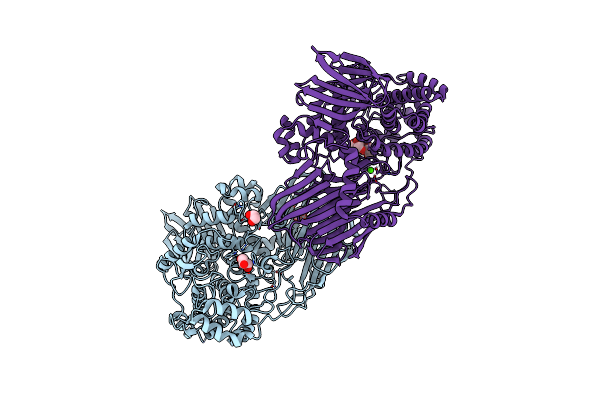
Crystal Structure Of The Gh74 Xyloglucanase From Xanthomonas Campestris (Xcc1752)
Organism: Xanthomonas campestris pv. campestris (strain atcc 33913 / dsm 3586 / ncppb 528 / lmg 568 / p 25)
Method: X-RAY DIFFRACTION Resolution:1.95 Å Release Date: 2021-05-26 Classification: HYDROLASE Ligands: EDO, NA, IOD |
 |
Crystal Structure Of The Gh31 Alpha-Xylosidase (Xac1773) From Xanthomonas Citri
Organism: Xanthomonas axonopodis pv. citri (strain 306)
Method: X-RAY DIFFRACTION Resolution:1.87 Å Release Date: 2021-05-26 Classification: HYDROLASE Ligands: K, XYS, GOL |
 |
Mycobacterium Tuberculosis Dihydrofolate Reductase In Complex With 5-Methyl-1-Phenyl-1H-Pyrazole-4-Carboxylic Acid (Fragment 1)
Organism: Mycobacterium tuberculosis
Method: X-RAY DIFFRACTION Resolution:1.76 Å Release Date: 2020-07-15 Classification: BIOSYNTHETIC PROTEIN Ligands: CO, 9FH, NDP, PO4 |
 |
Mycobacterium Tuberculosis Dihydrofolate Reductase In Complex With 3-(Furan-2-Yl)-1-Methyl-1H-Pyrazole-5-Carboxylic Acid (Fragment 2)
Organism: Mycobacterium tuberculosis
Method: X-RAY DIFFRACTION Resolution:1.85 Å Release Date: 2020-07-15 Classification: BIOSYNTHETIC PROTEIN Ligands: NDP, CO, RKV, PO4 |
 |
Mycobacterium Tuberculosis Dihydrofolate Reductase In Complex With Ethyl 2-Methyl Thiazole-4-Carboxylate(Fragment 3)
Organism: Mycobacterium tuberculosis
Method: X-RAY DIFFRACTION Resolution:1.83 Å Release Date: 2020-07-15 Classification: BIOSYNTHETIC PROTEIN Ligands: CO, NAP, PO4, RJY, SO4 |
 |
Mycobacterium Tuberculosis Dihydrofolate Reductase In Complex With 3-(Piperidin-1-Ylmethyl)Benzoic Acid(Fragment 11)
Organism: Mycobacterium tuberculosis (strain atcc 25177 / h37ra)
Method: X-RAY DIFFRACTION Resolution:1.84 Å Release Date: 2020-07-15 Classification: BIOSYNTHETIC PROTEIN Ligands: RK4, NAP, CO, PO4, GOL, SO4 |
 |
Mycobacterium Tuberculosis Dihydrofolate Reductase In Complex With 3-((Thiophen-2-Ylthio)Methyl)Benzoic Acid (Fragment 13)
Organism: Mycobacterium tuberculosis (strain atcc 25618 / h37rv)
Method: X-RAY DIFFRACTION Resolution:1.69 Å Release Date: 2020-07-15 Classification: BIOSYNTHETIC PROTEIN Ligands: JBB, NAP, CO, SO4, PO4 |
 |
Mycobacterium Tuberculosis Dihydrofolate Reductase In Complex With 3-(Phenoxymethyl)Benzoic Acid(Fragment 14)
Organism: Mycobacterium tuberculosis (strain atcc 25618 / h37rv)
Method: X-RAY DIFFRACTION Resolution:1.76 Å Release Date: 2020-07-15 Classification: BIOSYNTHETIC PROTEIN Ligands: CO, NAP, 4RG, PO4 |
 |
Mycobacterium Tuberculosis Dihydrofolate Reductase In Complex With 4-(3,4-Dihydro-2H-Benzo[B][1,4]Dioxepin-7-Yl)-4-Oxobutanoic Acid(Fragment 16)
Organism: Mycobacterium tuberculosis (strain atcc 25618 / h37rv)
Method: X-RAY DIFFRACTION Resolution:2.01 Å Release Date: 2020-07-15 Classification: BIOSYNTHETIC PROTEIN Ligands: RKY, CO, NAP, GOL, PO4 |
 |
Mycobacterium Tuberculosis Dihydrofolate Reductase In Complex With 4-(Trifluoromethyl)Benzene-1,2-Diamine(Fragment 17)
Organism: Mycobacterium tuberculosis (strain atcc 25618 / h37rv)
Method: X-RAY DIFFRACTION Resolution:2.30 Å Release Date: 2020-07-15 Classification: BIOSYNTHETIC PROTEIN Ligands: CO, X0V, GOL, NAP |
 |
Organism: Mycobacterium tuberculosis (strain atcc 25618 / h37rv)
Method: X-RAY DIFFRACTION Resolution:2.24 Å Release Date: 2020-07-15 Classification: BIOSYNTHETIC PROTEIN Ligands: CO, NAP, RPJ, SO4 |
 |
Organism: Mycobacterium tuberculosis (strain atcc 25177 / h37ra)
Method: X-RAY DIFFRACTION Resolution:2.00 Å Release Date: 2020-07-15 Classification: BIOSYNTHETIC PROTEIN Ligands: NDP, CO, RPM, PO4, SO4 |
 |
Organism: Mycobacterium tuberculosis (strain atcc 25177 / h37ra)
Method: X-RAY DIFFRACTION Resolution:2.68 Å Release Date: 2020-07-15 Classification: BIOSYNTHETIC PROTEIN Ligands: CO, NAP, RPV |