
Search Count: 57
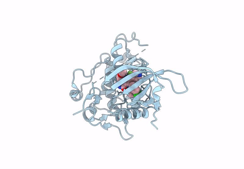 |
Organism: Homo sapiens
Method: X-RAY DIFFRACTION Resolution:2.50 Å Release Date: 2025-01-29 Classification: TRANSFERASE Ligands: A1IEZ |
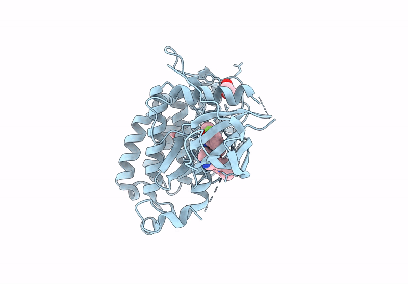 |
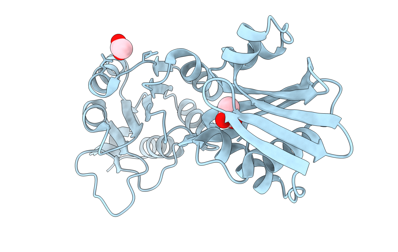
Organism: Homo sapiens
Method: X-RAY DIFFRACTION Resolution:1.78 Å Release Date: 2025-01-29 Classification: TRANSFERASE Ligands: EDO, A1IE0 |
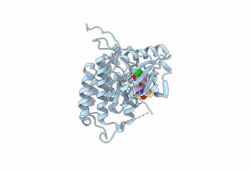 |
Organism: Homo sapiens
Method: X-RAY DIFFRACTION Resolution:2.06 Å Release Date: 2025-01-29 Classification: ONCOPROTEIN Ligands: DTT, A1IEZ, CL |
 |
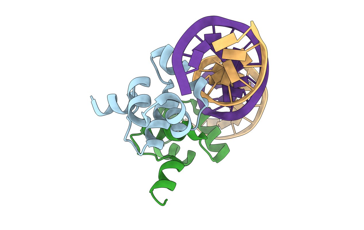
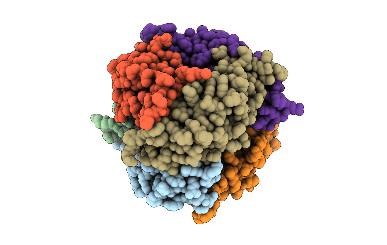
Organism: Homo sapiens, Synthetic construct
Method: ELECTRON MICROSCOPY Release Date: 2024-10-30 Classification: DNA BINDING PROTEIN/DNA |
 |
Organism: Homo sapiens, Synthetic construct
Method: ELECTRON MICROSCOPY Release Date: 2024-10-30 Classification: DNA BINDING PROTEIN/DNA |
 |
Organism: Homo sapiens, Synthetic construct
Method: ELECTRON MICROSCOPY Release Date: 2024-10-30 Classification: DNA BINDING PROTEIN/DNA |
 |
Organism: Homo sapiens, Synthetic construct
Method: ELECTRON MICROSCOPY Release Date: 2024-10-30 Classification: DNA BINDING PROTEIN/DNA |
 |
Organism: Escherichia phage n15, Enterobacteria phage n15
Method: X-RAY DIFFRACTION Resolution:1.60 Å Release Date: 2019-05-15 Classification: transcription/dna |
 |
Structure Of Phospholipase D Beta1B1I From Sicarius Terrosus Venom At 2.14 A Resolution
Organism: Sicarius terrosus
Method: X-RAY DIFFRACTION Resolution:2.14 Å Release Date: 2015-03-18 Classification: LYASE Ligands: MG |
 |
Organism: Escherichia coli
Method: X-RAY DIFFRACTION Resolution:1.60 Å Release Date: 2014-07-16 Classification: LYASE Ligands: ZN, ZSP, ACT |
 |
Organism: Escherichia coli
Method: X-RAY DIFFRACTION Resolution:2.05 Å Release Date: 2014-07-16 Classification: LYASE Ligands: ZN, 2K8 |
 |
Organism: Escherichia coli
Method: X-RAY DIFFRACTION Resolution:1.99 Å Release Date: 2014-07-16 Classification: LYASE Ligands: ZN, FMT |
 |
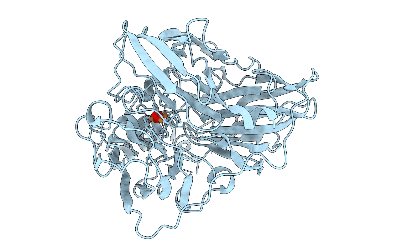
Organism: Archaeoglobus fulgidus
Method: X-RAY DIFFRACTION Resolution:1.59 Å Release Date: 2012-06-20 Classification: HYDROLASE Ligands: ACT |
 |
2.1A Crystal Structure Of The Phosphate Bound Atp Binding Domain Of Archaeoglobus Fulgidus Copb
Organism: Archaeoglobus fulgidus
Method: X-RAY DIFFRACTION Resolution:2.10 Å Release Date: 2012-06-20 Classification: HYDROLASE Ligands: PO4, MPO |
 |
Organism: Escherichia coli
Method: X-RAY DIFFRACTION Resolution:1.50 Å Release Date: 2012-02-29 Classification: OXIDOREDUCTASE Ligands: CU, CU1, O |
 |
Organism: Escherichia coli
Method: X-RAY DIFFRACTION Resolution:2.00 Å Release Date: 2011-10-19 Classification: OXIDOREDUCTASE Ligands: C2O, CU |
 |
Organism: Escherichia coli
Method: X-RAY DIFFRACTION Resolution:1.45 Å Release Date: 2011-10-19 Classification: OXIDOREDUCTASE Ligands: CU |
 |
Cueo At 1.1 A Resolution Including Residues In Previously Disordered Region
Organism: Escherichia coli
Method: X-RAY DIFFRACTION Resolution:1.10 Å Release Date: 2011-09-07 Classification: OXIDOREDUCTASE Ligands: CU, O, EDO |
 |
Organism: Escherichia coli
Method: X-RAY DIFFRACTION Resolution:1.50 Å Release Date: 2011-08-17 Classification: OXIDOREDUCTASE Ligands: CU, SO4, ACT |
 |
Organism: Escherichia coli
Method: X-RAY DIFFRACTION Resolution:2.00 Å Release Date: 2011-08-17 Classification: OXIDOREDUCTASE Ligands: CU, AG, O |