
Search Count: 728
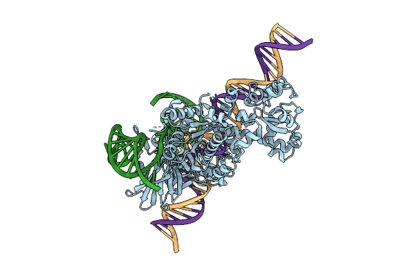 |
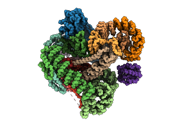
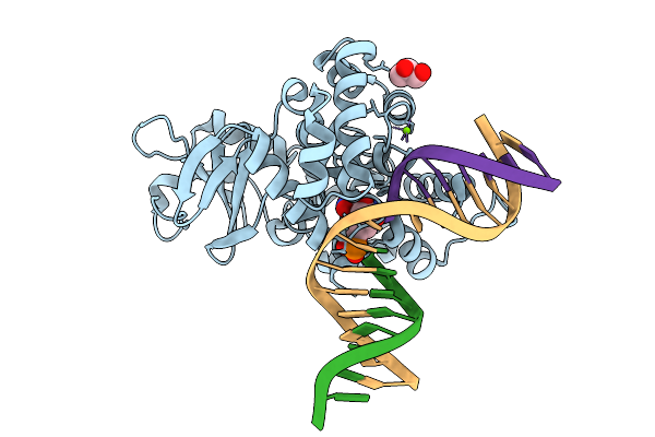
Cryo-Em Structure Of Esacas9_Nng-Guide Rna-Target Dna Complex In An Interrogation State
Organism: Staphylococcus aureus
Method: ELECTRON MICROSCOPY Release Date: 2025-11-05 Classification: DNA BINDING PROTEIN |
 |
Cryo-Em Structure Of Esacas9_Nng-Guide Rna-Target Dna Complex In An Interrogation State
Organism: Staphylococcus aureus
Method: ELECTRON MICROSCOPY Release Date: 2025-11-05 Classification: DNA BINDING PROTEIN |
 |
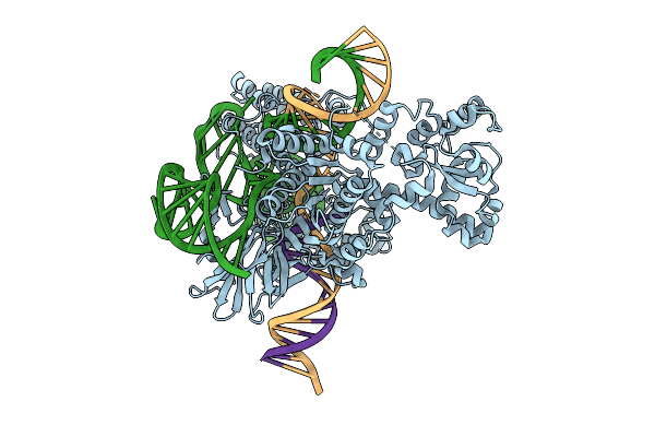
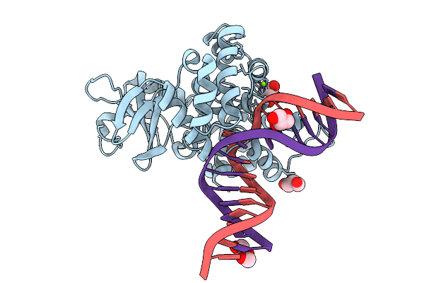
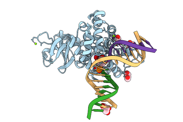
Cryo-Em Structure Of Esacas9_Nng-Guide Rna-Target Dna Complex In A Translocation State
Organism: Staphylococcus aureus
Method: ELECTRON MICROSCOPY Release Date: 2025-11-05 Classification: DNA BINDING PROTEIN Ligands: MG |
 |
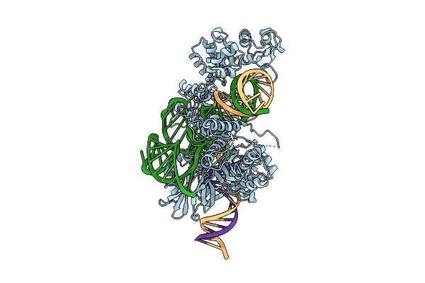
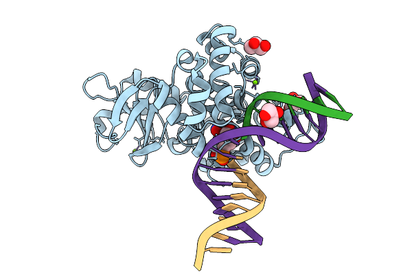
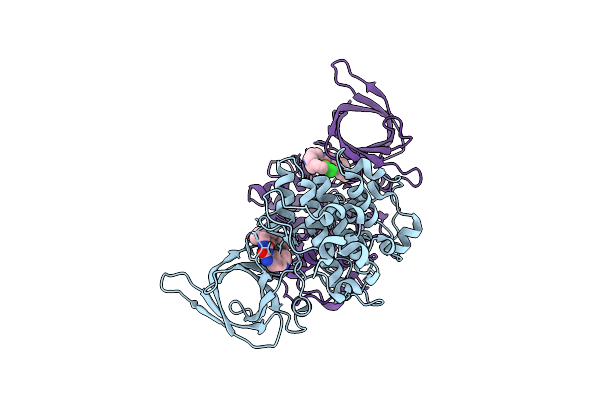
Cryo-Em Structure Of Esacas9_Nng-Guide Rna-Target Dna Complex In A Catalytically Active State
Organism: Staphylococcus aureus
Method: ELECTRON MICROSCOPY Release Date: 2025-11-05 Classification: DNA BINDING PROTEIN Ligands: MG |
 |
Crystal Structure Of The Mycobacterium Tuberculosis Regulator Virs (N-Terminal Fragment 4-208) In Complex With The Drug Candidate Alpibectir
Organism: Mycobacterium tuberculosis h37rv
Method: X-RAY DIFFRACTION Release Date: 2025-10-29 Classification: TRANSCRIPTION Ligands: YPQ |
 |
Human Carbonic Anhydrase Ii In Complex With Biguanide Derivative Inhibitor 1-Carbamimidamido-N-[2-(4-Sulfamoylphenyl)Ethyl]Methanimidamide
Organism: Homo sapiens
Method: X-RAY DIFFRACTION Release Date: 2025-10-29 Classification: LYASE Ligands: ZN, A1IRV, DMS |
 |
Human Carbonic Anhydrase Ii In Complex With B-Thujaplicin (2-Hydroxy-4-(Propan-2-Yl)Cyclohepta-2,4,6-Trien-1-One)
Organism: Homo sapiens
Method: X-RAY DIFFRACTION Release Date: 2025-10-29 Classification: LYASE Ligands: ZN, A1IRU |
 |
Organism: Homo sapiens, Mus musculus
Method: ELECTRON MICROSCOPY Release Date: 2025-10-22 Classification: STRUCTURAL PROTEIN |
 |
Crystal Structure Of M. Hassiacum Gpgs Co-Crystallized With Udp-Glucose (Ph 7.2)
Organism: Mycolicibacterium hassiacum dsm 44199
Method: X-RAY DIFFRACTION Release Date: 2025-09-24 Classification: TRANSFERASE Ligands: TLA, UDP |
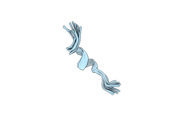 |
Organism: Rana temporaria
Method: SOLUTION NMR Release Date: 2025-09-17 Classification: ANTIMICROBIAL PROTEIN |
 |
High-Resolution X-Ray Structure Of Human Pc1/3 (Pcsk1) Prodomain R77A,R80A,R81A Triple-Mutant
|
 |
X-Ray Structure Of Furin (Pcsk3) In Complex With The Pc1/3 (Pcsk1) Prodomain Mutant R78K,R80A
Organism: Homo sapiens
Method: X-RAY DIFFRACTION Release Date: 2025-09-10 Classification: HYDROLASE Ligands: CA, NA |
 |
X-Ray Structure Of Furin (Pcsk3) In Complex With The Pc1/3 (Pcsk1) Prodomain Mutant R77A,R80A,R81A
Organism: Homo sapiens
Method: X-RAY DIFFRACTION Release Date: 2025-09-10 Classification: HYDROLASE Ligands: CA, NA, NAG |
 |
Crystal Structure Of Human 15-Pgdh In Complex With Small Molecule Compound 1
Organism: Homo sapiens
Method: X-RAY DIFFRACTION Release Date: 2025-09-10 Classification: OXIDOREDUCTASE/OXIDOREDUCTASE INHIBITOR Ligands: A1CH2, NAD, IOD, EDO, GOL |
 |
Crystal Structure Of Human 15-Pgdh In Complex With Small Molecule Compound 8B
Organism: Homo sapiens
Method: X-RAY DIFFRACTION Release Date: 2025-09-03 Classification: OXIDOREDUCTASE Ligands: NAD, SWL, EDO |
 |
Crystal Structure Of Human 8-Oxoguanine Glycosylase K249H Mutant Bound To The Substrate 8-Oxoguanine Dna At Ph 8.0 Under 277 K
Organism: Homo sapiens
Method: X-RAY DIFFRACTION Release Date: 2025-07-23 Classification: DNA/HYDROLASE Ligands: PEG, GOL, MG, NA |
 |
Crystal Structure Of Human 8-Oxoguanine Glycosylase K249H Mutant Bound To The Reaction Intermediate Derived From The Crystal Soaked Into The Solution At Ph 4.0 Under 277 K For 24 Hourss
Organism: Homo sapiens
Method: X-RAY DIFFRACTION Release Date: 2025-07-23 Classification: DNA/HYDROLASE Ligands: A1LXK, MG, GOL |
 |
Crystal Structure Of Human 8-Oxoguanine Glycosylase K249H Mutant Bound To The Reaction Intermediate Derived From The Crystal Soaked Into The Solution At Ph 4.0 Under 277 K For 2.5 Hours
Organism: Homo sapiens
Method: X-RAY DIFFRACTION Release Date: 2025-07-23 Classification: DNA/HYDROLASE Ligands: A1LXK, MG, GOL |
 |
Crystal Structure Of Human 8-Oxoguanine Glycosylase K249H Mutant Bound To The Reaction Intermediate Derived From The Crystal Soaked Into The Solution At Ph 4.0 Under 298 K For 3 Weeks
Organism: Homo sapiens
Method: X-RAY DIFFRACTION Release Date: 2025-07-23 Classification: DNA/HYDROLASE Ligands: A1LXK, GOL, MG, NA |
 |
Organism: Homo sapiens
Method: X-RAY DIFFRACTION Release Date: 2025-07-23 Classification: TRANSFERASE Ligands: A1IXL |