
Search Count: 32
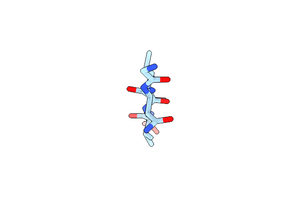 |
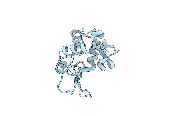
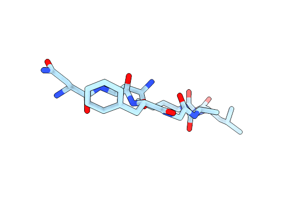
Structure Of The Amyloid Forming Peptide Gnlvs (Residues 26-30) From The Eosinophil Major Basic Protein (Embp)
Organism: Homo sapiens
Method: X-RAY DIFFRACTION Resolution:1.45 Å Release Date: 2015-03-18 Classification: PROTEIN FIBRIL |
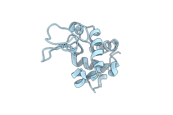 |
Organism: Gallus gallus
Method: X-RAY DIFFRACTION Resolution:2.60 Å Release Date: 2013-12-11 Classification: HYDROLASE |
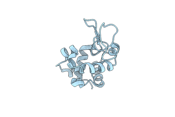 |
Organism: Gallus gallus
Method: X-RAY DIFFRACTION Resolution:2.00 Å Release Date: 2013-12-11 Classification: HYDROLASE |
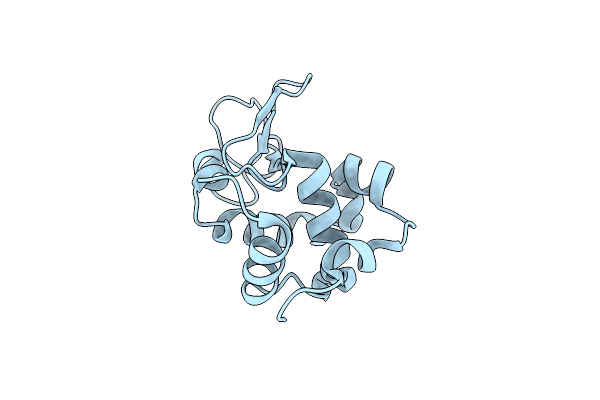 |
Laser-Induced Microfragmentation Of Lysozyme Crystals Allows X-Ray Nanodiffraction Characterization Of Individual Domains (Lb4)
Organism: Gallus gallus
Method: X-RAY DIFFRACTION Resolution:2.30 Å Release Date: 2013-11-20 Classification: HYDROLASE |
 |
Laser-Induced Microfragmentation Of Lysozyme Crystals Allows X-Ray Nanodiffraction Characterization Of Individual Domains (Lb5)
Organism: Gallus gallus
Method: X-RAY DIFFRACTION Resolution:2.50 Å Release Date: 2013-11-20 Classification: HYDROLASE |
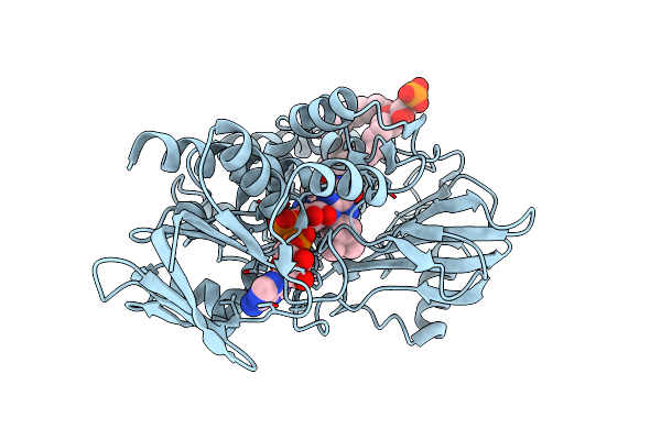 |
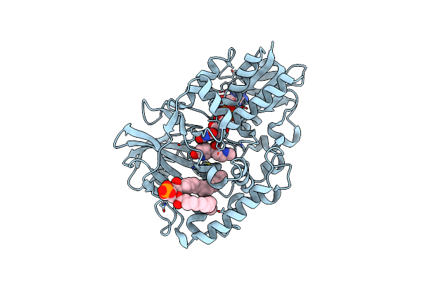
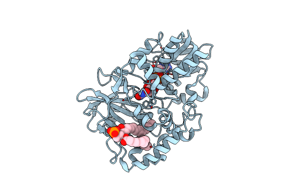
Organism: Arthrobacter nicotinovorans
Method: X-RAY DIFFRACTION Resolution:2.75 Å Release Date: 2011-06-15 Classification: OXIDOREDUCTASE Ligands: NCA, FAD, GP7 |
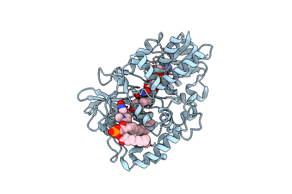 |
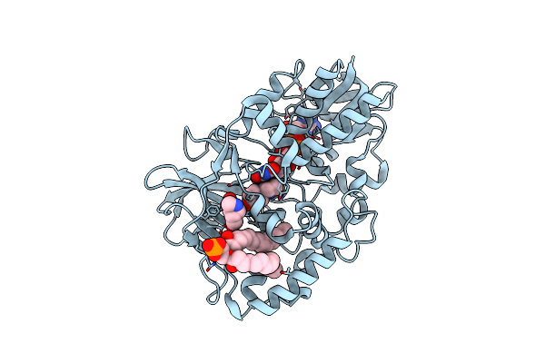
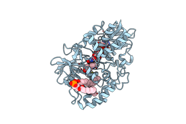
Complex Of 6-Hydroxy-L-Nicotine Oxidase With Intermediate Methylmyosmine Product Formed During Catalytic Turnover
Organism: Arthrobacter nicotinovorans
Method: X-RAY DIFFRACTION Resolution:2.25 Å Release Date: 2011-03-23 Classification: OXIDOREDUCTASE Ligands: HNL, FAD, GP7, HNH |
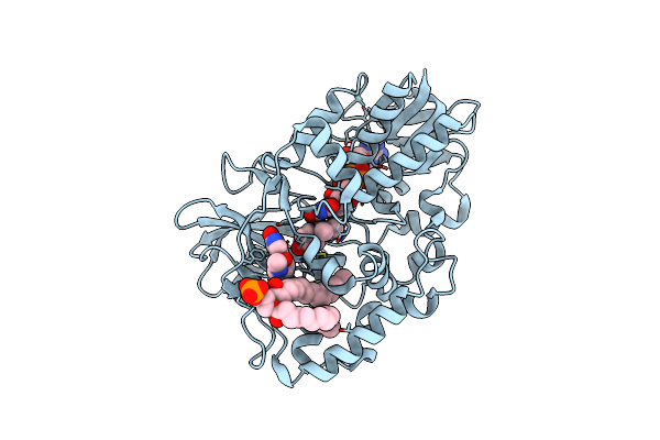 |
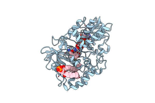
Complex Of 6-Hydroxy-L-Nicotine Oxidase With Final Ketone Product Formed During Catalytic Turnover
Organism: Arthrobacter nicotinovorans
Method: X-RAY DIFFRACTION Resolution:2.10 Å Release Date: 2011-03-23 Classification: OXIDOREDUCTASE Ligands: HNL, FAD, GP7, HNM |
 |
Organism: Arthrobacter nicotinovorans
Method: X-RAY DIFFRACTION Resolution:2.85 Å Release Date: 2011-03-23 Classification: OXIDOREDUCTASE Ligands: HNM, FAD, GP7 |
 |
Complex Of 6-Hydroxy-L-Nicotine Oxidase With Inhibitor Bound At Active Site And Turnover Product At Exit Cavity
Organism: Arthrobacter nicotinovorans
Method: X-RAY DIFFRACTION Resolution:2.15 Å Release Date: 2011-03-23 Classification: OXIDOREDUCTASE Ligands: HNK, FAD, GP7, HNM |
 |
Organism: Arthrobacter nicotinovorans
Method: X-RAY DIFFRACTION Resolution:2.20 Å Release Date: 2011-03-23 Classification: OXIDOREDUCTASE Ligands: SRO, FAD, GP7 |
 |
Organism: Arthrobacter nicotinovorans
Method: X-RAY DIFFRACTION Resolution:2.65 Å Release Date: 2011-03-23 Classification: OXIDOREDUCTASE Ligands: LDP, FAD, GP7 |
 |
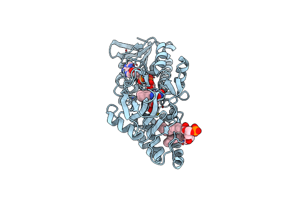
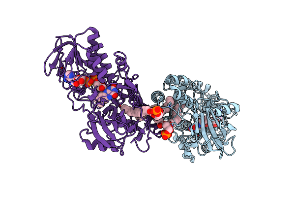
Crystal Structure Of Inhibitor-Bound In Active Centre 6-Hydroxy-L-Nicotine Oxidase From Arthrobacter Nicotinovorans
Organism: Arthrobacter nicotinovorans
Method: X-RAY DIFFRACTION Resolution:2.19 Å Release Date: 2011-03-23 Classification: OXIDOREDUCTASE/OXIDOREDUCTASE INHIBITOR Ligands: HNK, FB0, GP7 |
 |
Crystal Structure Of 6-Hydroxy-L-Nicotine Oxidase From Arthrobacter Nicotinovorans
Organism: Arthrobacter nicotinovorans
Method: X-RAY DIFFRACTION Resolution:1.95 Å Release Date: 2010-01-19 Classification: OXIDOREDUCTASE Ligands: FAD, GP7 |
 |
Crystal Structure Of Substrate-Bound 6-Hydroxy-L-Nicotine Oxidase From Arthrobacter Nicotinovorans
Organism: Arthrobacter nicotinovorans
Method: X-RAY DIFFRACTION Resolution:2.05 Å Release Date: 2010-01-19 Classification: OXIDOREDUCTASE Ligands: HNL, FAD, GP7 |
 |
Crystal Structure Of Apo-Form 6-Hydroxy-L-Nicotine Oxidase, Crystal Form P3121
Organism: Arthrobacter nicotinovorans
Method: X-RAY DIFFRACTION Resolution:2.85 Å Release Date: 2010-01-19 Classification: OXIDOREDUCTASE Ligands: FAD, GP7 |
 |
 |
Method: X-RAY DIFFRACTION
Resolution:1.80 Å Release Date: 2008-07-01 Classification: PROTEIN FIBRIL Ligands: HOH |
 |
Organism: Trichoderma longibrachiatum
Method: X-RAY DIFFRACTION Resolution:1.50 Å Release Date: 2008-05-13 Classification: HYDROLASE |
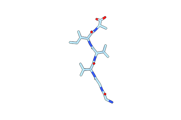 |
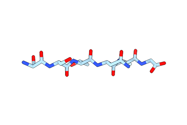
Crystal Structure Of The Amyloid-Fibril Forming Peptide Ggvvia Derived From The Alzheimer'S Amyloid Abeta (Abeta37-42).
|