
Search Count: 11
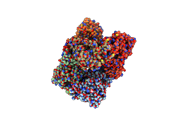 |
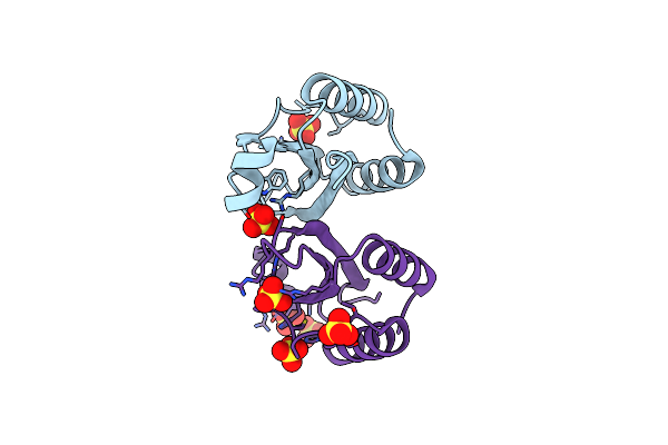
Organism: Cyanobium sp. pcc 7001
Method: X-RAY DIFFRACTION Resolution:2.30 Å Release Date: 2023-08-09 Classification: PHOTOSYNTHESIS Ligands: ZN, GOL, SO4, EDO, RUB, CO2, BCT |
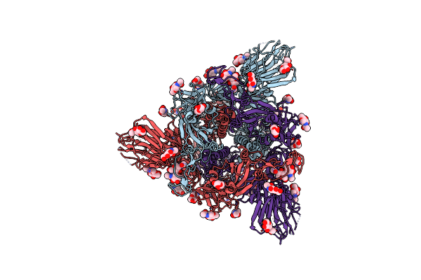 |
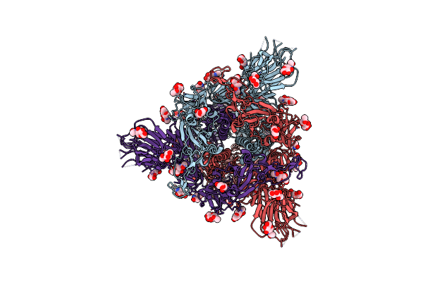
Structure Of Disulphide-Stabilized Sars-Cov-2 Spike Protein Trimer (X2 Disulphide-Bond Mutant, G413C, V987C, Single Arg S1/S2 Cleavage Site)
Organism: Severe acute respiratory syndrome coronavirus 2
Method: ELECTRON MICROSCOPY Release Date: 2020-07-22 Classification: VIRAL PROTEIN Ligands: NAG |
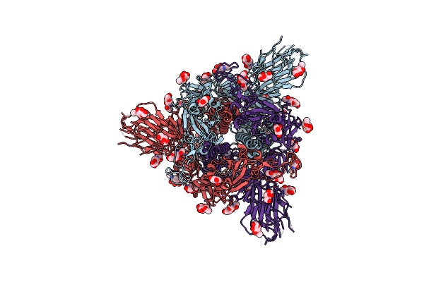 |
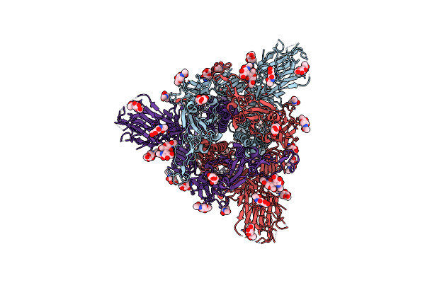
Structure Of Disulphide-Stabilized Sars-Cov-2 Spike Protein Trimer (X1 Disulphide-Bond Mutant, S383C, D985C, K986P, V987P, Single Arg S1/S2 Cleavage Site) In Closed State
Organism: Severe acute respiratory syndrome coronavirus 2
Method: ELECTRON MICROSCOPY Release Date: 2020-07-22 Classification: VIRAL PROTEIN Ligands: NAG |
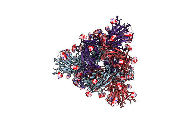 |
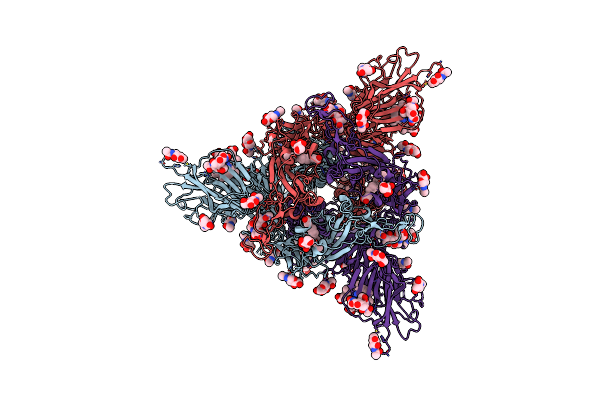
Structure Of Disulphide-Stabilized Sars-Cov-2 Spike Protein Trimer (X1 Disulphide-Bond Mutant, S383C, D985C, K986P, V987P, Single Arg S1/S2 Cleavage Site) In Locked State
Organism: Severe acute respiratory syndrome coronavirus 2
Method: ELECTRON MICROSCOPY Release Date: 2020-07-22 Classification: VIRAL PROTEIN Ligands: NAG, BLA |
 |
Structure Of Sars-Cov-2 Spike Protein Trimer (Single Arg S1/S2 Cleavage Site) In Closed State
Organism: Severe acute respiratory syndrome coronavirus 2
Method: ELECTRON MICROSCOPY Release Date: 2020-07-22 Classification: VIRAL PROTEIN Ligands: NAG |
 |
Structure Of Sars-Cov-2 Spike Protein Trimer (K986P, V987P, Single Arg S1/S2 Cleavage Site) In Closed State
Organism: Severe acute respiratory syndrome coronavirus 2
Method: ELECTRON MICROSCOPY Release Date: 2020-07-22 Classification: VIRAL PROTEIN Ligands: NAG |
 |
Structure Of Sars-Cov-2 Spike Protein Trimer (K986P, V987P, Single Arg S1/S2 Cleavage Site) In Locked State
Organism: Severe acute respiratory syndrome coronavirus 2
Method: ELECTRON MICROSCOPY Release Date: 2020-07-22 Classification: VIRAL PROTEIN Ligands: BLA, NAG, EIC |
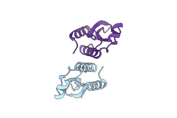 |
Crystal Structure Of The Small Subunit-Like Domain 1 Of Ccmm From Synechococcus Elongatus (Strain Pcc 7942), Thiol-Oxidized Form
Organism: Synechococcus elongatus (strain pcc 7942)
Method: X-RAY DIFFRACTION Resolution:1.65 Å Release Date: 2018-12-12 Classification: PROTEIN BINDING |
 |
Crystal Structure Of The Small Subunit-Like Domain 1 Of Ccmm From Synechococcus Elongatus (Strain Pcc 7942)
Organism: Synechococcus elongatus (strain pcc 7942)
Method: X-RAY DIFFRACTION Resolution:1.20 Å Release Date: 2018-12-12 Classification: PROTEIN BINDING Ligands: SO4 |
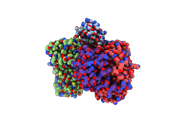 |
Structure Of The Repeat Unit In The Network Formed By Ccmm And Rubisco From Synechococcus Elongatus
Organism: Synechococcus elongatus (strain pcc 7942)
Method: ELECTRON MICROSCOPY Release Date: 2018-12-12 Classification: PROTEIN BINDING |
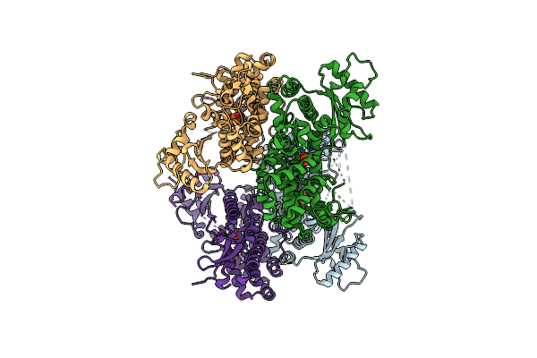 |
Organism: Homo sapiens
Method: X-RAY DIFFRACTION Resolution:3.10 Å Release Date: 2011-11-16 Classification: HYDROLASE Ligands: PO4, ZN |