
Search Count: 1,725
All
Selected
 |
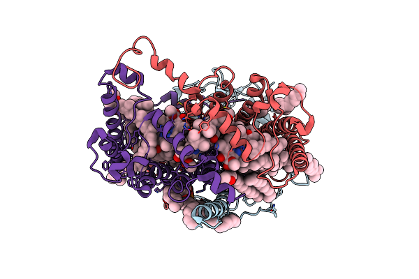
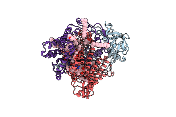
F(M197)H Mutant Structure Of Photosynthetic Reaction Center From Rhodobacter Sphaeroides Strain Rv By Fixed-Target Serial Synchrotron Crystallography (100K, 26Kev)
Organism: Rhodobacter sphaeroides
Method: X-RAY DIFFRACTION Resolution:1.73 Å Release Date: 2022-07-13 Classification: PHOTOSYNTHESIS Ligands: LDA, UNL, OLC, BCL, BPH, NKP, U10, FE, SPN, EDO |
 |
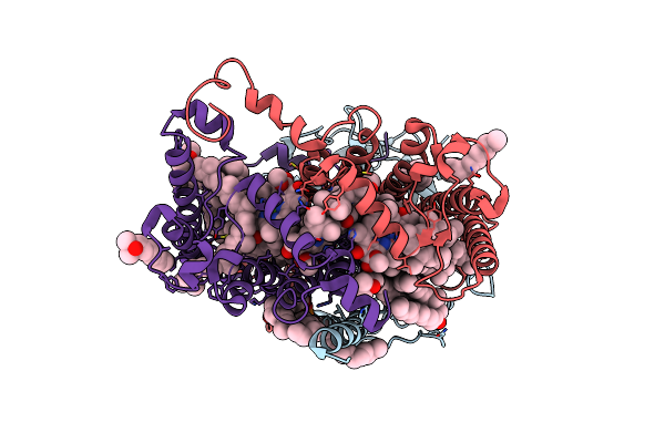
F(M197)H Mutant Structure Of Photosynthetic Reaction Center From Rhodobacter Sphaeroides Strain Rv By Fixed-Target Serial Synchrotron Crystallography (Room Temperature, 12Kev)
Organism: Rhodobacter sphaeroides
Method: X-RAY DIFFRACTION Resolution:2.22 Å Release Date: 2022-07-13 Classification: PHOTOSYNTHESIS Ligands: MYS, LDA, OLC, BCL, BPH, NKP, U10, SPN, FE, EDO |
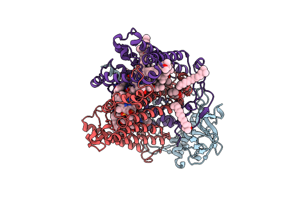 |
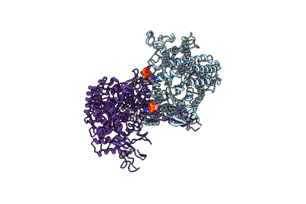
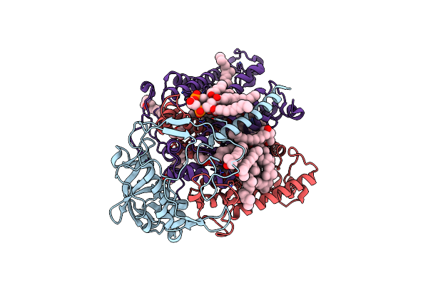
Crystal Structure Of R. Sphaeroides Photosynthetic Reaction Center Variant; W252V Mutant
Organism: Rhodobacter sphaeroides
Method: X-RAY DIFFRACTION Resolution:2.79 Å Release Date: 2022-04-27 Classification: ELECTRON TRANSPORT Ligands: BCL, BPH, U10, CL, LDA, FE, SPO, CDL |
 |
F(M197)H Mutant Structure Of Photosynthetic Reaction Center From Rhodobacter Sphaeroides Strain Rv Lsp Crystallization
Organism: Rhodobacter sphaeroides
Method: X-RAY DIFFRACTION Resolution:2.10 Å Release Date: 2022-04-27 Classification: PHOTOSYNTHESIS Ligands: LDA, UNL, OLC, BCL, BPH, NKP, FE, U10, SPN, EDO |
 |
F(M197)H Mutant Structure Of Photosynthetic Reaction Center From Rhodobacter Sphaeroides Strain Rv By Fixed-Target Serial Synchrotron Crystallography (Room Temperature, 26Kev)
Organism: Rhodobacter sphaeroides (strain atcc 17023 / dsm 158 / jcm 6121 / nbrc 12203 / ncimb 8253 / ath 2.4.1.)
Method: X-RAY DIFFRACTION Resolution:2.04 Å Release Date: 2022-04-27 Classification: PHOTOSYNTHESIS Ligands: LDA, OLC, MYS, BCL, BPH, NKP, U10, FE, SPN, EDO, PO4 |
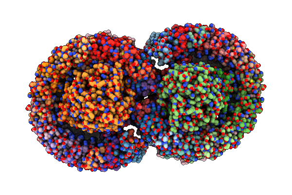 |
Organism: Rhodobacter sphaeroides 2.4.1
Method: ELECTRON MICROSCOPY Release Date: 2022-04-27 Classification: PHOTOSYNTHESIS Ligands: BCL, BPB, U10, PC1, FE2, SPO, CDL |
 |
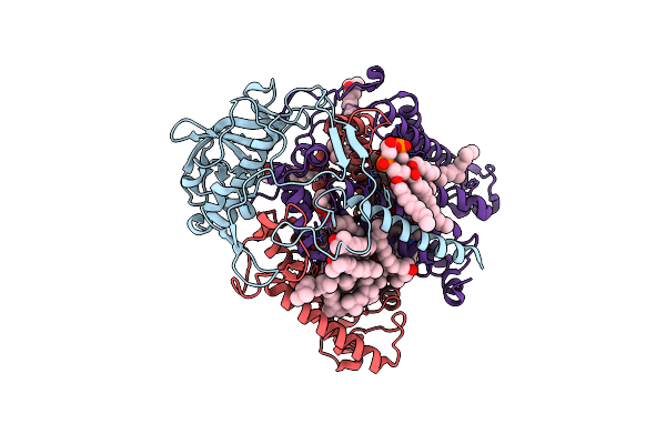
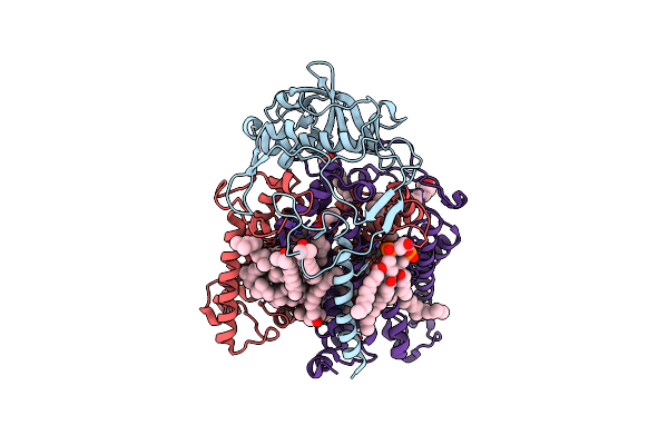
Structure Of Photosynthetic Lh1-Rc Super-Complex Of Rhodobacter Sphaeroides Dimer
Organism: Rhodobacter sphaeroides f. sp. denitrificans
Method: ELECTRON MICROSCOPY Release Date: 2022-04-27 Classification: PHOTOSYNTHESIS Ligands: BCL, BPH, U10, LMT, PGV, FE, SPO, CDL, PTY, LDA |
 |
Structure Of Photosynthetic Lh1-Rc Super-Complex Of Rhodobacter Sphaeroides Lacking Protein-U
Organism: Rhodobacter sphaeroides f. sp. denitrificans
Method: ELECTRON MICROSCOPY Release Date: 2022-04-27 Classification: PHOTOSYNTHESIS Ligands: BCL, BPH, U10, PGV, LDA, LMT, FE, SPO, CDL, PTY |
 |
Organism: Rhodobacter sphaeroides (strain atcc 17023 / 2.4.1 / ncib 8253 / dsm 158)
Method: X-RAY DIFFRACTION Resolution:2.65 Å Release Date: 2022-01-12 Classification: OXIDOREDUCTASE Ligands: DTP, MG |
 |
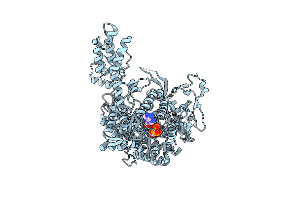
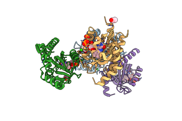
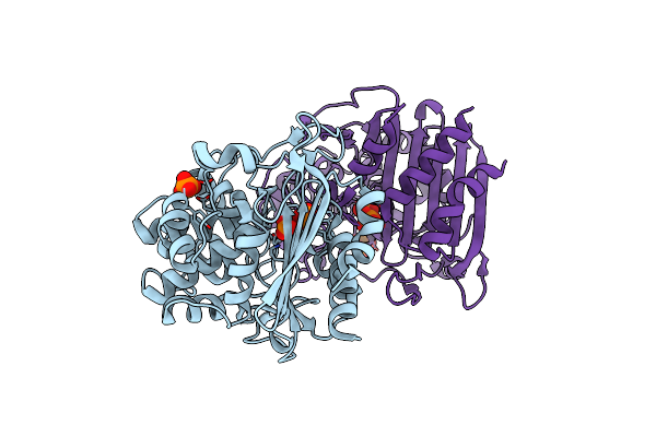
The Serp Optimized Structure Of Ribonucleotide Reductase From Rhodobacter Sphaeroides
Organism: Rhodobacter sphaeroides
Method: X-RAY DIFFRACTION Resolution:2.78 Å Release Date: 2022-01-12 Classification: OXIDOREDUCTASE Ligands: DTP, MG |
 |
Crystal Structure Of R. Sphaeroides Photosynthetic Reaction Center Variant; Y(M210)3-Chlorotyrosine
Organism: Rhodobacter sphaeroides
Method: X-RAY DIFFRACTION Resolution:2.30 Å Release Date: 2021-12-29 Classification: PHOTOSYNTHESIS Ligands: LDA, BCL, BPH, U10, CL, FE, SPO, CDL |
 |
Crystal Structure Of R. Sphaeroides Photosynthetic Reaction Center Variant; Y(M210)3-Bromotyrosine
Organism: Rhodobacter sphaeroides
Method: X-RAY DIFFRACTION Resolution:2.48 Å Release Date: 2021-12-29 Classification: PHOTOSYNTHESIS Ligands: LDA, BCL, BPH, U10, CL, FE, SPO, CDL |
 |
Crystal Structure Of R. Sphaeroides Photosynthetic Reaction Center Variant; Y(M210)3-Iodotyrosine
Organism: Rhodobacter sphaeroides
Method: X-RAY DIFFRACTION Resolution:2.85 Å Release Date: 2021-12-29 Classification: PHOTOSYNTHESIS Ligands: LDA, BPH, U10, BCL, FE, SPO, CDL |
 |
Crystal Structure Of R. Sphaeroides Photosynthetic Reaction Center Variant; Y(M210)3-Methyltyrosine
Organism: Rhodobacter sphaeroides
Method: X-RAY DIFFRACTION Resolution:2.75 Å Release Date: 2021-12-29 Classification: PHOTOSYNTHESIS Ligands: LDA, BCL, BPH, U10, FE, SPO, CDL |
 |
Crystal Structure Of R. Sphaeroides Photosynthetic Reaction Center Variant; Y(M210)3-Nitrotyrosine
Organism: Rhodobacter sphaeroides
Method: X-RAY DIFFRACTION Resolution:3.10 Å Release Date: 2021-12-29 Classification: PHOTOSYNTHESIS Ligands: LDA, BCL, BPH, U10, CL, FE, SPO, CDL |
 |
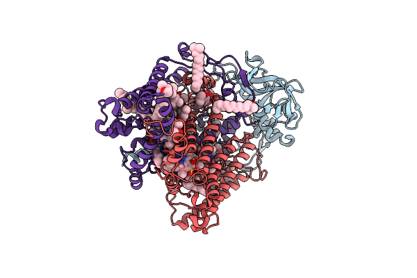
Structure Of Photosynthetic Lh1-Rc Super-Complex Of Rhodobacter Sphaeroides Monomer
Organism: Rhodobacter sphaeroides
Method: ELECTRON MICROSCOPY Release Date: 2021-11-10 Classification: PHOTOSYNTHESIS Ligands: BCL, BPH, U10, PGV, LDA, FE, SPO, LMT, CDL |
 |
Cryo-Em Structure Of The Rhodobacter Sphaeroides Rc-Lh1-Pufxy Monomer Complex At 2.5 A
Organism: Rhodobacter sphaeroides (strain atcc 17023 / dsm 158 / jcm 6121 / nbrc 12203 / ncimb 8253 / ath 2.4.1.), Rhodobacter sphaeroides (strain atcc 17023 / 2.4.1 / ncib 8253 / dsm 158)
Method: ELECTRON MICROSCOPY Release Date: 2021-10-13 Classification: PHOTOSYNTHESIS Ligands: BCL, SPO, LMT, 3PE, BPH, U10, UQ1, CD4, FE |
 |
Organism: Rhodobacter sphaeroides (strain atcc 17025 / ath 2.4.3)
Method: X-RAY DIFFRACTION Resolution:1.90 Å Release Date: 2021-06-30 Classification: SIGNALING PROTEIN Ligands: CA, FMN, ACT, MG, NA |
 |
Organism: Rhodobacter sphaeroides (strain atcc 17025 / ath 2.4.3)
Method: X-RAY DIFFRACTION Resolution:2.00 Å Release Date: 2021-06-30 Classification: SIGNALING PROTEIN Ligands: FMN, CL, SPD, NA |
 |
Organism: Rhodobacter sphaeroides 2.4.1
Method: X-RAY DIFFRACTION Resolution:2.21 Å Release Date: 2021-05-19 Classification: HYDROLASE Ligands: PO4 |