
Search Count: 18
 |
Organism: Loligo pealei, Todarodes pacificus
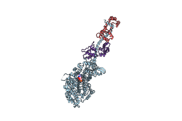
Method: X-RAY DIFFRACTION Resolution:3.10 Å Release Date: 2009-08-04 Classification: CONTRACTILE PROTEIN Ligands: MG, ADP |
 |
Organism: Loligo pealei, Todarodes pacificus
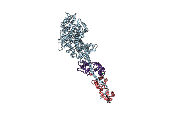
Method: X-RAY DIFFRACTION Resolution:2.60 Å Release Date: 2009-07-28 Classification: CONTRACTILE PROTEIN Ligands: MLI, CA |
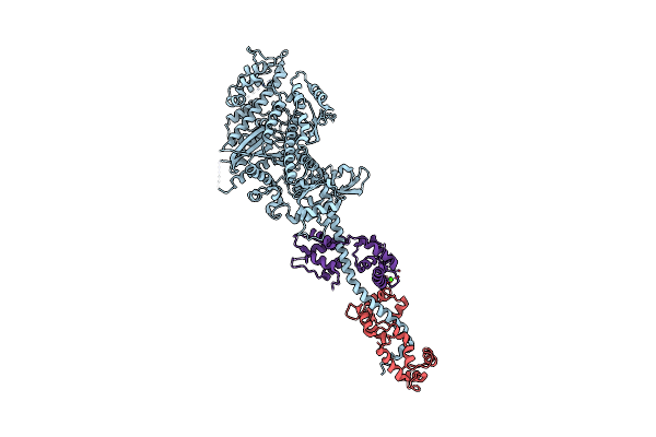 |
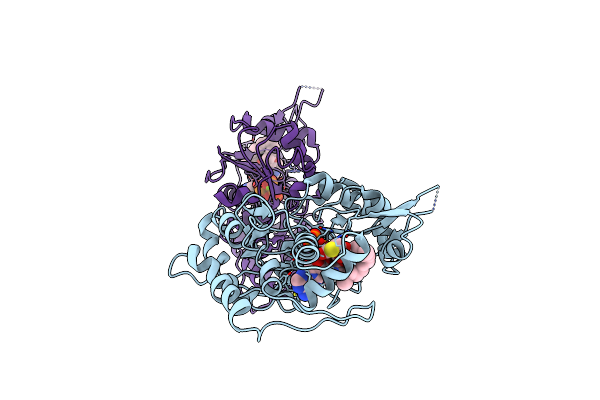
The Crystal Structure Of Rigor Like Squid Myosin S1 In The Absence Of Nucleotide
Organism: Loligo pealei, Todarodes pacificus
Method: X-RAY DIFFRACTION Resolution:3.40 Å Release Date: 2009-07-28 Classification: CONTRACTILE PROTEIN Ligands: CA |
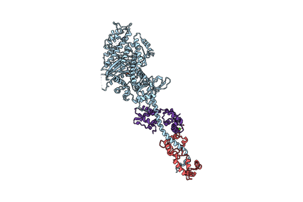 |
Organism: Loligo pealei, Todarodes pacificus
Method: X-RAY DIFFRACTION Resolution:3.30 Å Release Date: 2009-07-28 Classification: CONTRACTILE PROTEIN Ligands: SO4, CA |
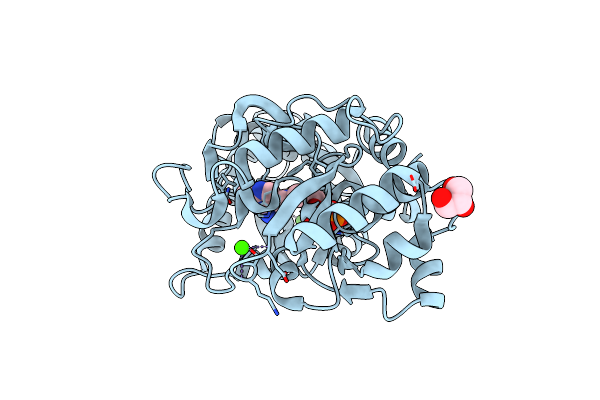 |
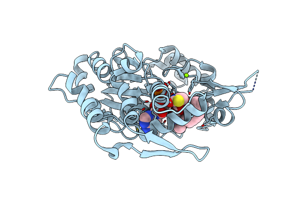
Structural Insights Of The Mycoplasma Hyorhinis Protein Mh-P37: A Putative Thiamine Pyrophosphate Transporter
Organism: Mycoplasma hyorhinis
Method: X-RAY DIFFRACTION Resolution:1.60 Å Release Date: 2009-06-23 Classification: TPP BINDING PROTEIN Ligands: TPP, GOL, CA, BR |
 |
Structure Determination Of The Cancer-Associated Mycoplasma Hyorhinis Protein Mh-P37
Organism: Mycoplasma hyorhinis
Method: X-RAY DIFFRACTION Resolution:1.90 Å Release Date: 2008-10-21 Classification: TPP Binding Protein Ligands: TPP, CA, CL |
 |
Structure Determination Of The Cancer-Associated Mycoplasma Hyorhinis Protein Mh-P37
Organism: Mycoplasma hyorhinis
Method: X-RAY DIFFRACTION Resolution:1.90 Å Release Date: 2008-10-21 Classification: TPP Binding Protein Ligands: I3C, TPP, CA, CL |
 |
Organism: Placopecten magellanicus
Method: X-RAY DIFFRACTION Resolution:3.25 Å Release Date: 2008-02-26 Classification: CONTRACTILE PROTEIN Ligands: CA |
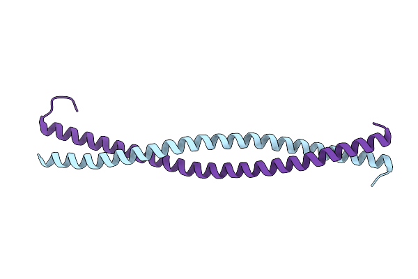 |
Crystal Structure Of The N-Terminal Region Of The Scallop Myosin Rod, Monoclinic (C2) Form
Organism: Argopecten irradians, Saccharomyces cerevisiae
Method: X-RAY DIFFRACTION Resolution:2.30 Å Release Date: 2008-01-08 Classification: CONTRACTILE PROTEIN Ligands: IOD |
 |
Crystal Structure Of The N-Terminal Region Of The Scallop Myosin Rod, Monoclinic (P21) Form
Organism: Argopecten irradians, Saccharomyces cerevisiae
Method: X-RAY DIFFRACTION Resolution:2.30 Å Release Date: 2008-01-08 Classification: CONTRACTILE PROTEIN |
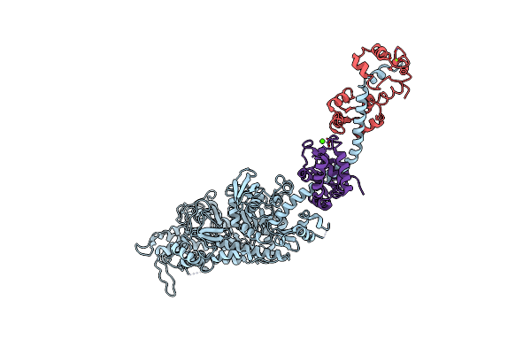 |
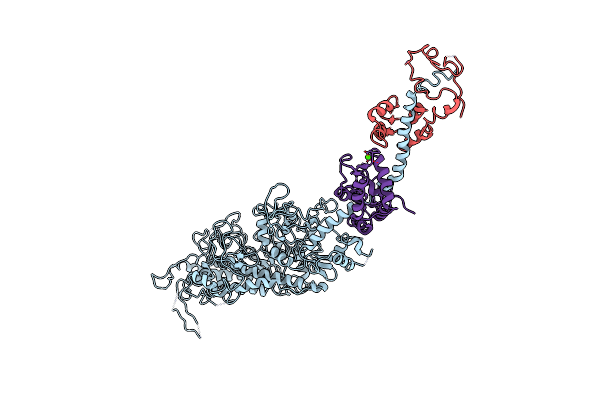
Rigor-Like Structures Of Muscle Myosins Reveal Key Mechanical Elements In The Transduction Pathways Of This Allosteric Motor
Organism: Placopecten magellanicus
Method: X-RAY DIFFRACTION Resolution:3.27 Å Release Date: 2007-05-29 Classification: CONTRACTILE PROTEIN Ligands: MG, CA |
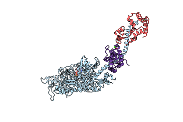 |
Rigor-Like Structures Of Muscle Myosins Reveal Key Mechanical Elements In The Transduction Pathways Of This Allosteric Motor
Organism: Placopecten magellanicus
Method: X-RAY DIFFRACTION Resolution:3.12 Å Release Date: 2007-05-29 Classification: CONTRACTILE PROTEIN Ligands: MG, ADP, CA |
 |
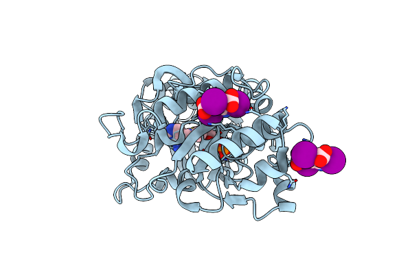
Organism: Homo sapiens
Method: X-RAY DIFFRACTION Resolution:2.40 Å Release Date: 2006-07-04 Classification: OXIDOREDUCTASE Ligands: MN, K |
 |
Organism: Homo sapiens
Method: X-RAY DIFFRACTION Resolution:2.40 Å Release Date: 2006-07-04 Classification: OXIDOREDUCTASE Ligands: MN, K |
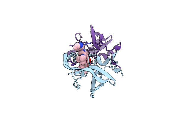 |
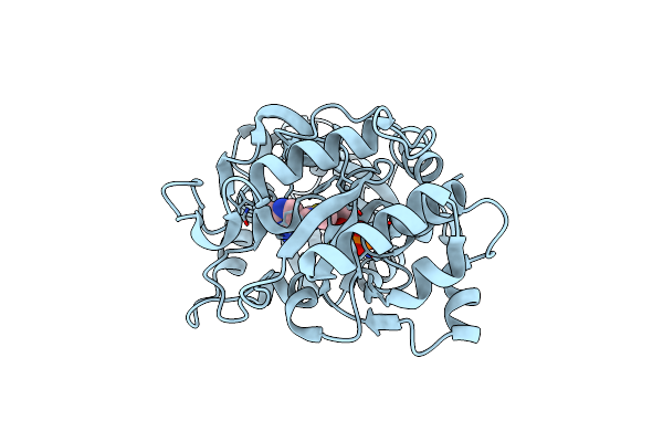
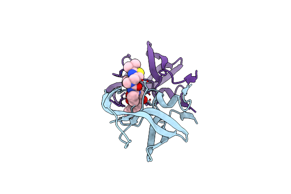
Comparing The Accumulation Of Active Site And Non-Active Site Mutations In The Hiv-1 Protease
Organism: Human immunodeficiency virus 1
Method: X-RAY DIFFRACTION Resolution:1.90 Å Release Date: 2004-10-05 Classification: HYDROLASE Ligands: MK1 |
 |
Comparing The Accumulation Of Active Site And Non-Active Site Mutations In The Hiv-1 Protease
Organism: Human immunodeficiency virus 1
Method: X-RAY DIFFRACTION Resolution:2.50 Å Release Date: 2004-10-05 Classification: HYDROLASE/HYDROLASE INHIBITOR Ligands: RIT |
 |
Actin Crystal Dynamics: Structural Implications For F-Actin Nucleation, Polymerization And Branching Mediated By The Anti-Parallel Dimer
Organism: Oryctolagus cuniculus
Method: X-RAY DIFFRACTION Resolution:2.30 Å Release Date: 2003-12-16 Classification: STRUCTURAL PROTEIN Ligands: MG, ATP, LAR |
 |
Actin Crystal Dynamics: Structural Implications For F-Actin Nucleation, Polymerization And Branching Mediated By The Anti-Parallel Dimer
Organism: Oryctolagus cuniculus
Method: X-RAY DIFFRACTION Release Date: 2003-12-16 Classification: STRUCTURAL PROTEIN Ligands: MG, ATP, LAR |