
Search Count: 19
 |
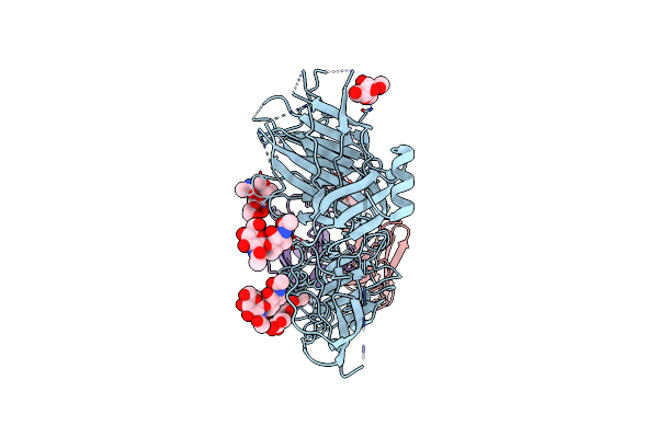
Organism: Severe acute respiratory syndrome coronavirus 2, Tequatrovirus t4, Homo sapiens
Method: ELECTRON MICROSCOPY Release Date: 2024-01-31 Classification: VIRAL PROTEIN |
 |
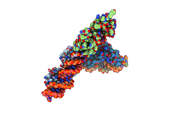
Organism: Severe acute respiratory syndrome coronavirus 2, Enterobacteria phage t4, Homo sapiens
Method: ELECTRON MICROSCOPY Release Date: 2024-01-31 Classification: VIRAL PROTEIN Ligands: NAG |
 |
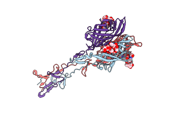
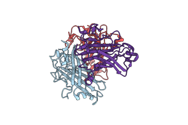
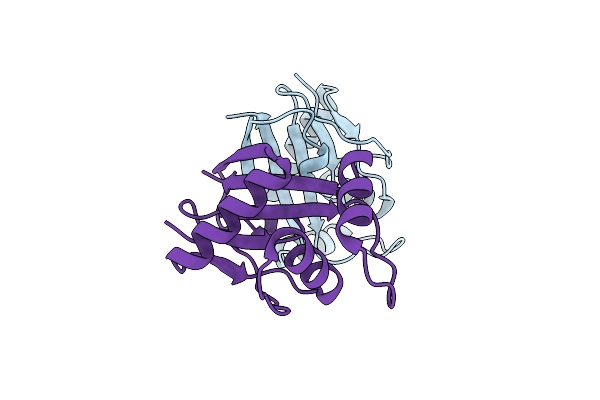
Structure Of The Plasmodium Falciparum Sip2 Dna-Binding Ap2 Tandem Repeat In Complex With Two Spe2 Half-Sites
Organism: Plasmodium falciparum (isolate 3d7), Plasmodium falciparum 3d7
Method: X-RAY DIFFRACTION Resolution:3.10 Å Release Date: 2020-10-07 Classification: DNA BINDING PROTEIN |
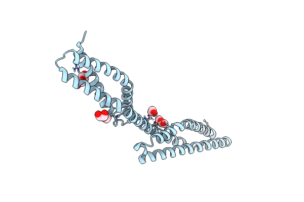 |
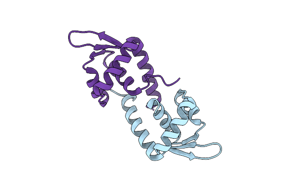
Organism: Homo sapiens
Method: X-RAY DIFFRACTION Resolution:1.54 Å Release Date: 2017-04-12 Classification: STRUCTURAL PROTEIN Ligands: EDO, PG4 |
 |
Crystal Structure Of The T1L Reovirus Attachment Protein Sigma1 In Complex With The Gm2 Glycan
Organism: Mammalian orthoreovirus 1
Method: X-RAY DIFFRACTION Resolution:3.60 Å Release Date: 2012-12-05 Classification: VIRAL PROTEIN |
 |
Crystal Structure Of The T1L Reovirus Attachment Protein Sigma1 In Complex With Alpha-2,3-Sialyllactose
Organism: Mammalian orthoreovirus 1
Method: X-RAY DIFFRACTION Resolution:3.50 Å Release Date: 2012-12-05 Classification: VIRAL PROTEIN |
 |
Structure Of Reovirus Attachment Protein Sigma1 In Complex With Alpha-2,3-Sialyllactose
Organism: Reovirus type 3
Method: X-RAY DIFFRACTION Resolution:2.25 Å Release Date: 2011-11-23 Classification: VIRAL PROTEIN |
 |
Structure Of Reovirus Attachment Protein Sigma1 In Complex With Alpha-2,6-Sialyllactose
Organism: Reovirus type 3
Method: X-RAY DIFFRACTION Resolution:2.79 Å Release Date: 2011-11-23 Classification: VIRAL PROTEIN Ligands: SIA |
 |
Structure Of Reovirus Attachment Protein Sigma1 In Complex With Alpha-2,8-Disialyllactose
Organism: Reovirus type 3
Method: X-RAY DIFFRACTION Resolution:2.28 Å Release Date: 2011-11-23 Classification: VIRAL PROTEIN |
 |
Organism: Human adenovirus b
Method: X-RAY DIFFRACTION Resolution:1.45 Å Release Date: 2008-11-25 Classification: VIRAL PROTEIN |
 |
Organism: Human adenovirus 7
Method: X-RAY DIFFRACTION Resolution:1.75 Å Release Date: 2008-11-25 Classification: VIRAL PROTEIN |
 |
Organism: Human adenovirus 14
Method: X-RAY DIFFRACTION Resolution:1.80 Å Release Date: 2008-11-25 Classification: VIRAL PROTEIN Ligands: GOL, IMD |
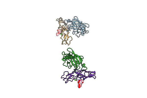 |
Human Adenovirus Type 11 Knob In Complex With Domains Scr1 And Scr2 Of Cd46 (Membrane Cofactor Protein, Mcp)
Organism: Human adenovirus 11p, Homo sapiens
Method: X-RAY DIFFRACTION Resolution:2.85 Å Release Date: 2007-01-09 Classification: VIRAL PROTEIN/immune system Ligands: CA |
 |
Organism: Sulfolobus turreted icosahedral virus
Method: X-RAY DIFFRACTION Resolution:2.39 Å Release Date: 2006-11-02 Classification: VIRAL PROTEIN |
 |
Organism: Sulfolobus turreted icosahedral virus
Method: X-RAY DIFFRACTION Resolution:2.20 Å Release Date: 2006-05-31 Classification: VIRAL PROTEIN/WINGED HELIX |
 |
Organism: Sulfolobus turreted icosahedral virus
Method: X-RAY DIFFRACTION Resolution:1.86 Å Release Date: 2005-09-29 Classification: VIRAL PROTEIN/TRANSFERASE Ligands: DIO, SO4, NI |
 |
Molecular Structure Of An Apolipoprotein Determined At 2.5-Angstroms Resolution
Organism: Locusta migratoria
Method: X-RAY DIFFRACTION Resolution:2.70 Å Release Date: 1994-01-31 Classification: LIPOPROTEIN |
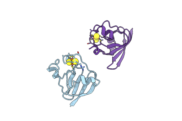 |
Crystallization And Structure Determination To 2.5-Angstroms Resolution Of The Oxidized [2Fe-2S] Ferredoxin Isolated From Anabaena 7120
Organism: Nostoc sp.
Method: X-RAY DIFFRACTION Resolution:2.50 Å Release Date: 1992-07-15 Classification: ELECTRON TRANSPORT Ligands: FES |
 |
The Molecular Structure Of The High Potential Iron-Sulfur Protein Isolated From Ectothiorhodospira Halophila Determined At 2.5-Angstroms Resolution
Organism: Halorhodospira halophila
Method: X-RAY DIFFRACTION Resolution:2.50 Å Release Date: 1992-07-15 Classification: ELECTRON TRANSFER (IRON-SULFUR PROTEIN) Ligands: SF4 |