
Search Count: 11
 |
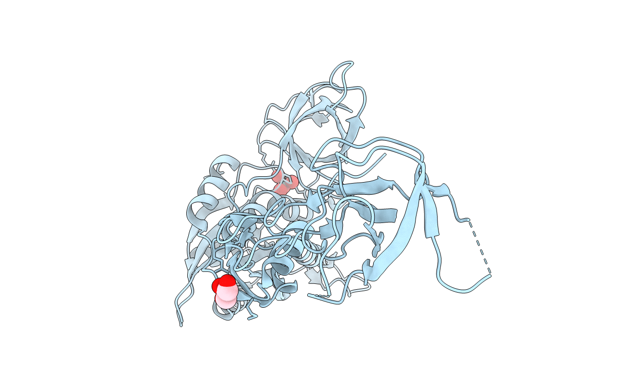
Organism: Pseudomonas aeruginosa ucbpp-pa14
Method: X-RAY DIFFRACTION Resolution:2.09 Å Release Date: 2018-08-01 Classification: OXIDOREDUCTASE |
 |
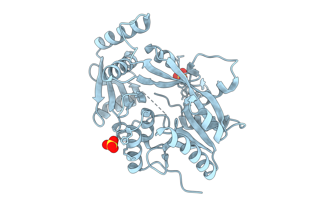
Crystal Structure Of The Pyoverdine Maturation Protein Pvdp In Complex With The Mock Substrates L-Tyrosine And Zinc.
Organism: Pseudomonas aeruginosa (strain ucbpp-pa14)
Method: X-RAY DIFFRACTION Resolution:2.70 Å Release Date: 2018-08-01 Classification: OXIDOREDUCTASE Ligands: ZN, TYR |
 |
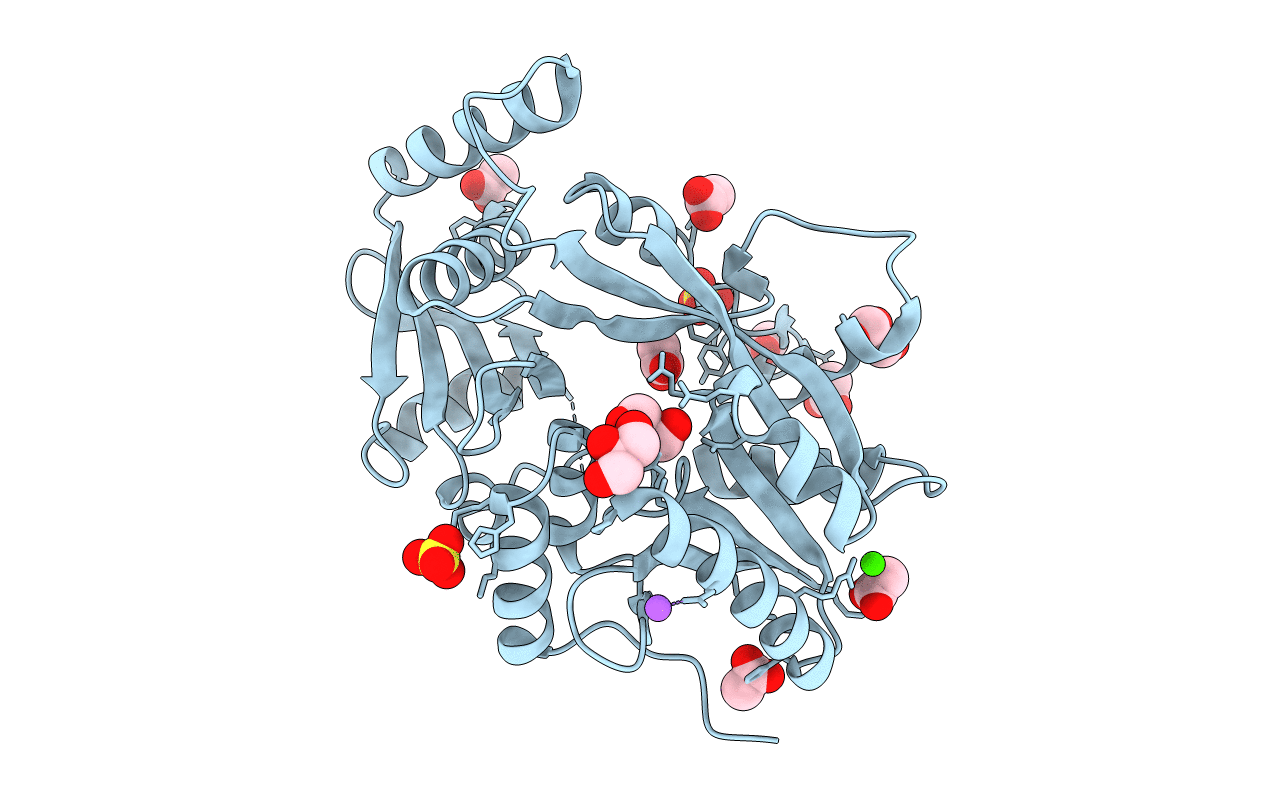
Crystal Structure Of The Mo-Insertase Domain Cnx1E From Arabidopsis Thaliana
Organism: Arabidopsis thaliana
Method: X-RAY DIFFRACTION Resolution:2.45 Å Release Date: 2017-02-15 Classification: TRANSFERASE Ligands: SO4, GOL, MG |
 |
Crystal Structure Of The Mo-Insertase Domain Cnx1E From Arabidopsis Thaliana In Complex With Molybdate
Organism: Arabidopsis thaliana
Method: X-RAY DIFFRACTION Resolution:2.84 Å Release Date: 2017-02-15 Classification: TRANSFERASE Ligands: MOO, GOL, MG |
 |
Organism: Myxococcus xanthus (strain dk 1622)
Method: X-RAY DIFFRACTION Resolution:2.05 Å Release Date: 2016-06-22 Classification: LYASE |
 |
Organism: Myxococcus xanthus (strain dk 1622)
Method: X-RAY DIFFRACTION Resolution:1.10 Å Release Date: 2016-06-22 Classification: LYASE Ligands: COA, MLI |
 |
Organism: Pseudomonas aeruginosa
Method: X-RAY DIFFRACTION Resolution:3.10 Å Release Date: 2015-08-19 Classification: TRANSFERASE Ligands: CL, SO4 |
 |
Organism: Pseudomonas aeruginosa
Method: X-RAY DIFFRACTION Resolution:2.30 Å Release Date: 2015-08-19 Classification: TRANSFERASE Ligands: SO4, GOL, ACT, CA, NA, CL |
 |
Organism: Bacillus licheniformis
Method: X-RAY DIFFRACTION Resolution:2.10 Å Release Date: 2015-08-19 Classification: TRANSFERASE Ligands: LYN, TRS |
 |
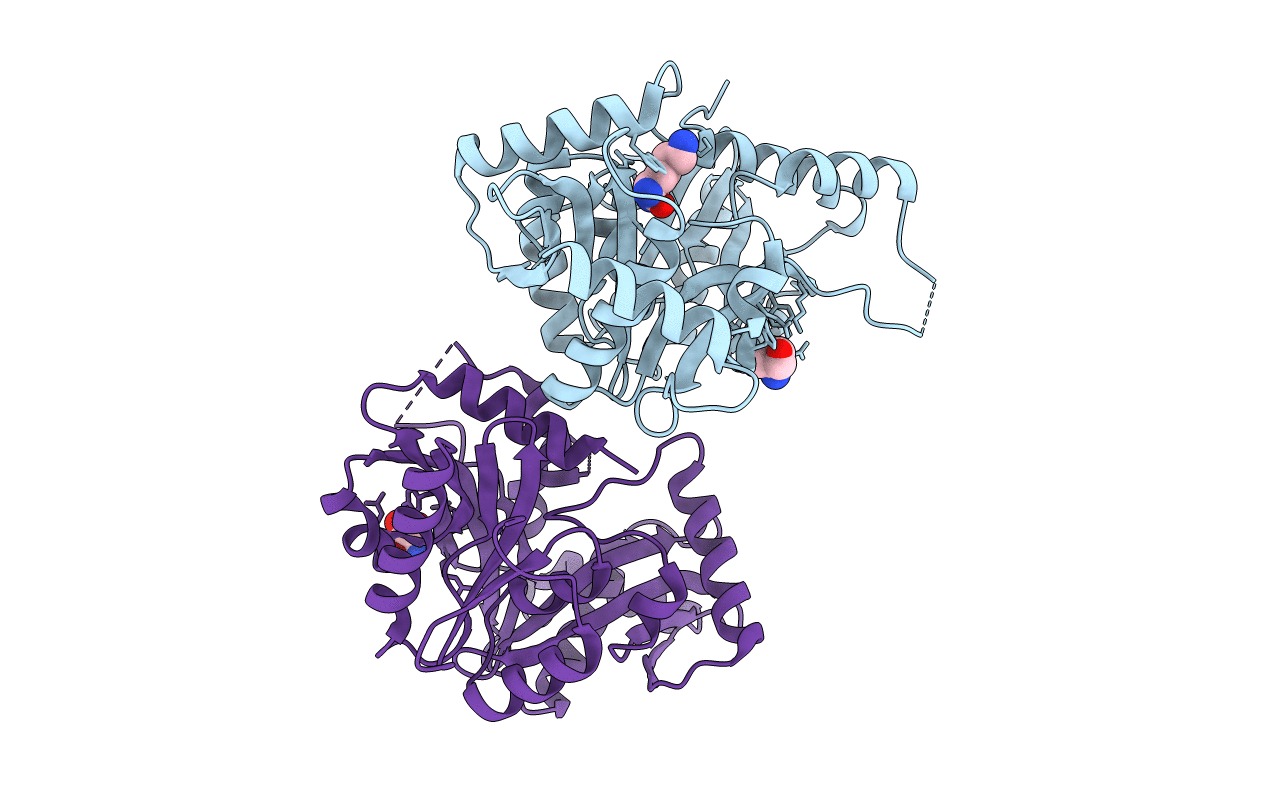
Crystal Structure Of The I-Bar Domain Of Irsp53 (Baiap2) In Complex With An Ehec Derived Tir Peptide
Organism: Homo sapiens, Escherichia coli o157\:h7
Method: X-RAY DIFFRACTION Resolution:2.11 Å Release Date: 2011-09-07 Classification: SIGNALING PROTEIN Ligands: SO4 |
 |
Functional Domain Of Inlj From Listeria Monocytogenes Includes A Cysteine Ladder
Organism: Listeria monocytogenes
Method: X-RAY DIFFRACTION Resolution:2.70 Å Release Date: 2008-06-17 Classification: CELL ADHESION Ligands: SO4, CL |