
Search Count: 18
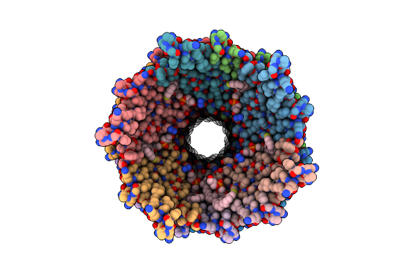 |
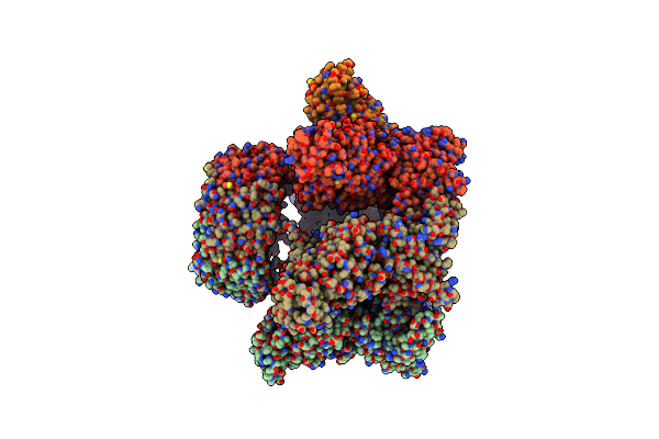
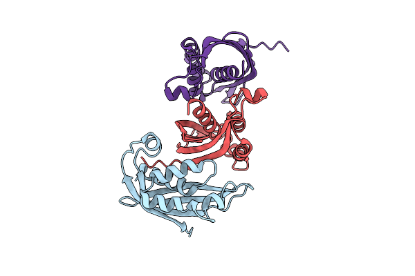
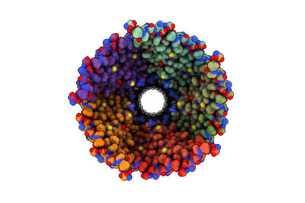
Cryo-Em Structure Of The R388 Plasmid Conjugative Pilus Reveals A Helical Polymer Characterised By An Unusual Pilin/Phospholipid Binary Complex
Organism: Escherichia coli hb101
Method: ELECTRON MICROSCOPY Release Date: 2024-07-24 Classification: PROTEIN FIBRIL Ligands: LHG |
 |
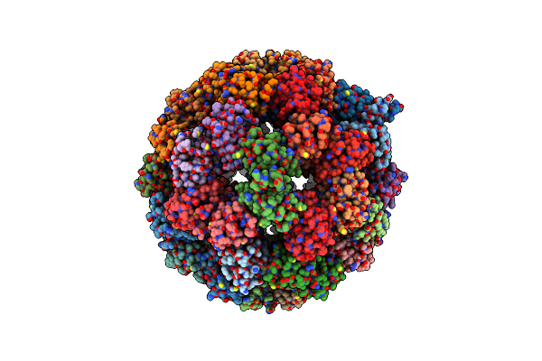
O-Layer Structure (Trwh/Virb7, Trwf/Virb9Ctd, Trwe/Virb10Ctd) Of The Outer Membrane Core Complex From The Fully-Assembled R388 Type Iv Secretion System Determined By Cryo-Em.
Organism: Escherichia coli
Method: ELECTRON MICROSCOPY Release Date: 2022-06-22 Classification: MEMBRANE PROTEIN |
 |
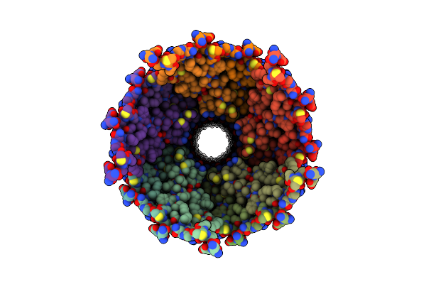
I-Layer Structure (Trwf/Virb9Ntd, Trwe/Virb10Ntd) Of The Outer Membrane Core Complex From The Fully-Assembled R388 Type Iv Secretion System Determined By Cryo-Em.
Organism: Escherichia coli
Method: ELECTRON MICROSCOPY Release Date: 2022-06-22 Classification: MEMBRANE PROTEIN |
 |
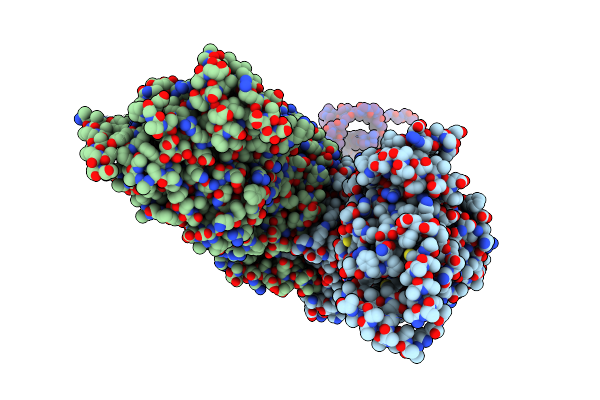
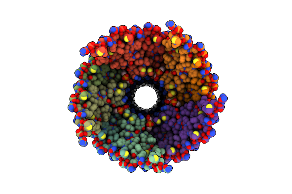
Stalk Complex Structure (Trwj/Virb5-Trwi/Virb6) From The Fully-Assembled R388 Type Iv Secretion System Determined By Cryo-Em.
Organism: Escherichia coli
Method: ELECTRON MICROSCOPY Release Date: 2022-06-22 Classification: MEMBRANE PROTEIN |
 |
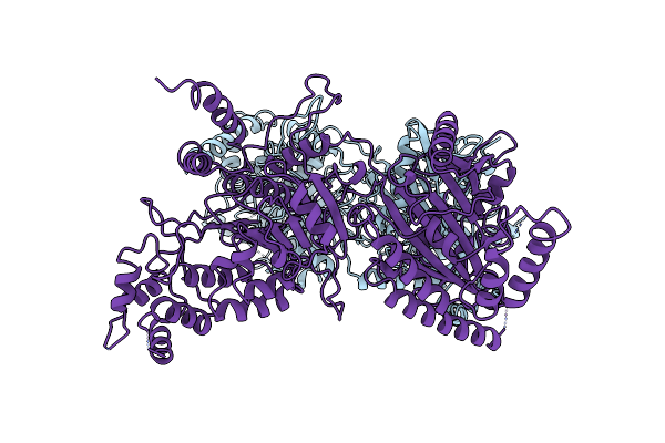
Hexameric Composite Model Of The Inner Membrane Complex (Imc) With The Arches From The Fully-Assembled R388 Type Iv Secretion System Determined By Cryo-Em.
Organism: Escherichia coli
Method: ELECTRON MICROSCOPY Release Date: 2022-06-22 Classification: MEMBRANE PROTEIN |
 |
Trwk/Virb4Unbound Trimer Of Dimers Complex (With Hcp1) From The R388 Type Iv Secretion System Determined By Cryo-Em.
Organism: Salmonella dublin, Pseudomonas aeruginosa (strain atcc 15692 / dsm 22644 / cip 104116 / jcm 14847 / lmg 12228 / 1c / prs 101 / pao1)
Method: ELECTRON MICROSCOPY Release Date: 2022-06-22 Classification: MEMBRANE PROTEIN |
 |
Trwk/Virb4Unbound Dimer Complex From R388 Type Iv Secretion System Determined By Cryo-Em.
Organism: Escherichia coli
Method: ELECTRON MICROSCOPY Release Date: 2022-06-22 Classification: MEMBRANE PROTEIN |
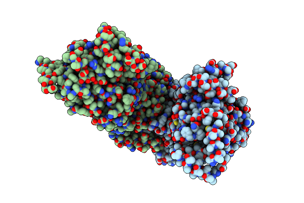 |
Inner Membrane Complex (Imc) Protomer Structure (Trwm/Virb3, Trwk/Virb4, Trwg/Virb8Tails) From The Fully-Assembled R388 Type Iv Secretion System Determined By Cryo-Em.
Organism: Escherichia coli
Method: ELECTRON MICROSCOPY Release Date: 2022-06-22 Classification: MEMBRANE PROTEIN |
 |
Arches Protomer (Trimer Of Trwg/Virb8Peri) Structure From The Fully-Assembled R388 Type Iv Secretion System Determined By Cryo-Em.
Organism: Escherichia coli
Method: ELECTRON MICROSCOPY Release Date: 2022-06-22 Classification: MEMBRANE PROTEIN |
 |
Organism: Legionella pneumophila
Method: ELECTRON MICROSCOPY Release Date: 2020-05-13 Classification: MEMBRANE PROTEIN Ligands: ZN |
 |
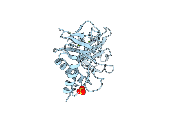
Fab Fragment Of An Antibody That Inhibits Polymerisation Of Alpha-1-Antitrypsin
Organism: Mus musculus
Method: X-RAY DIFFRACTION Resolution:1.90 Å Release Date: 2020-03-18 Classification: PROTEIN BINDING Ligands: SO4, NA, GOL |
 |
Organism: Listeria monocytogenes egd-e
Method: ELECTRON MICROSCOPY Release Date: 2019-08-21 Classification: ANTIMICROBIAL PROTEIN |
 |
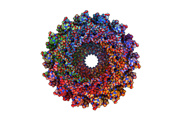
The Cryo-Electron Microscopy Structure Of The Type 1 Chaperone-Usher Pilus Rod
Organism: Escherichia coli j96
Method: ELECTRON MICROSCOPY Release Date: 2017-11-22 Classification: PROTEIN FIBRIL |
 |
Organism: Escherichia coli
Method: ELECTRON MICROSCOPY Release Date: 2016-11-02 Classification: PROTEIN FIBRIL Ligands: 6V6 |
 |
Organism: Escherichia coli
Method: ELECTRON MICROSCOPY Release Date: 2016-11-02 Classification: PROTEIN FIBRIL Ligands: 6V6 |
 |
Organism: Salmonella enterica subsp. enterica serovar typhi
Method: ELECTRON MICROSCOPY Release Date: 2016-09-28 Classification: PROTEIN FIBRIL Ligands: LHG |
 |
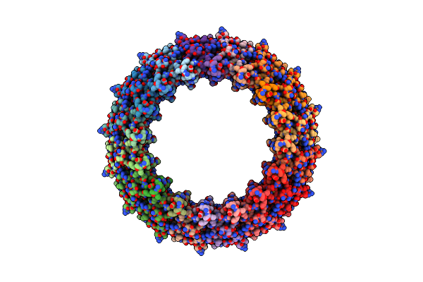
Structure Of A Chaperone-Usher Pilus Reveals The Molecular Basis Of Rod Uncoilin
Organism: Escherichia coli
Method: ELECTRON MICROSCOPY Resolution:3.80 Å Release Date: 2016-01-13 Classification: STRUCTURAL PROTEIN |
 |
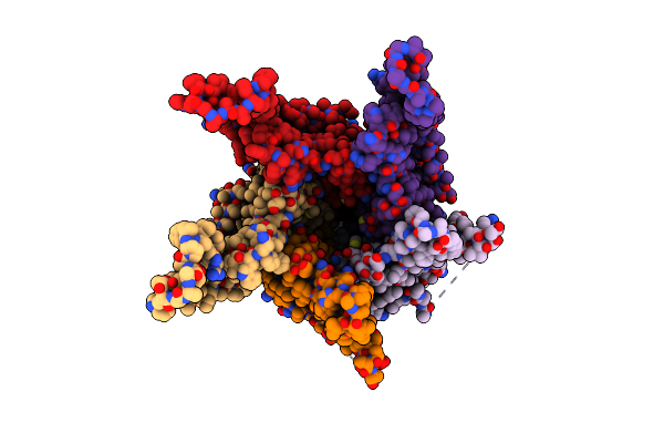
Structure Of A Translocation Signal Domain Mediating Conjugative Transfer By Type Iv Secretion Systems
Organism: Escherichia coli
Method: X-RAY DIFFRACTION Resolution:1.85 Å Release Date: 2013-06-19 Classification: HYDROLASE Ligands: SO4, MG |