
Search Count: 639
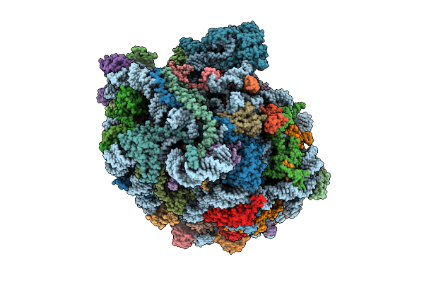 |
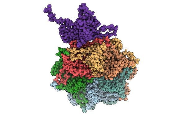
High Resolution Structure Of The Thermophilic 60S Ribosomal Subunit Of Chaetomium Thermophilum
Organism: Thermochaetoides thermophila dsm 1495
Method: ELECTRON MICROSCOPY Release Date: 2025-12-17 Classification: RIBOSOME Ligands: SPD, SPM, MG, K, ZN |
 |
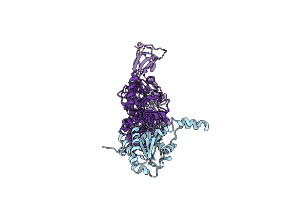
Organism: Saccharomyces cerevisiae
Method: X-RAY DIFFRACTION Release Date: 2025-11-05 Classification: DNA BINDING PROTEIN/DNA Ligands: ZN |
 |
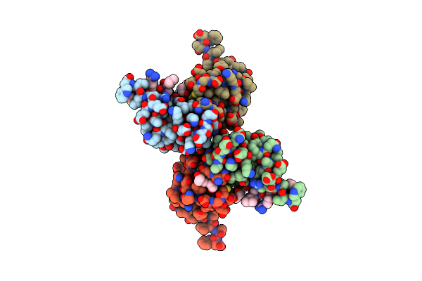
Organism: Homo sapiens, Synthetic construct, Sus scrofa
Method: X-RAY DIFFRACTION Release Date: 2025-11-05 Classification: MOTOR PROTEIN Ligands: GTP, MG, EDO, GDP, ANP |
 |
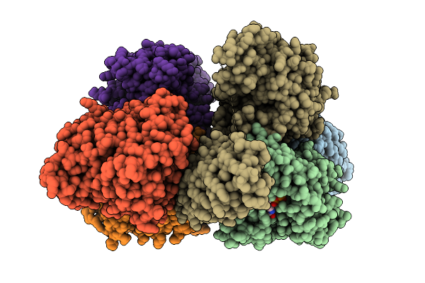
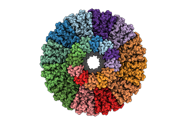
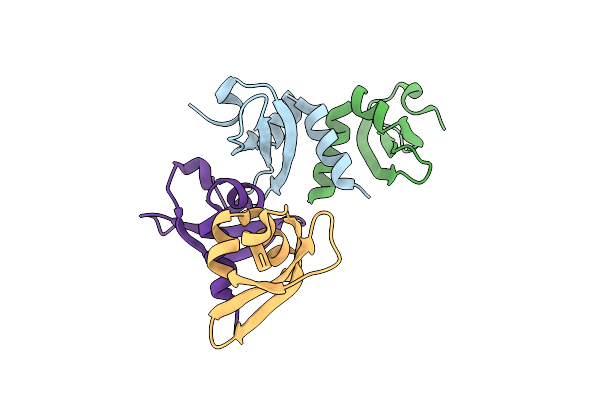
Cryo-Em Structure Of The Multimeric Phosphoenolpyruvate Binding Domain Of Staphylothermus Marinus Phosphoenolpyruvate Synthase
Organism: Staphylothermus marinus f1
Method: ELECTRON MICROSCOPY Release Date: 2025-10-29 Classification: CYTOSOLIC PROTEIN Ligands: MG |
 |
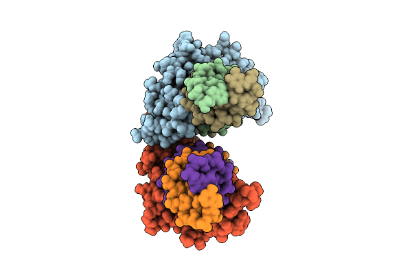
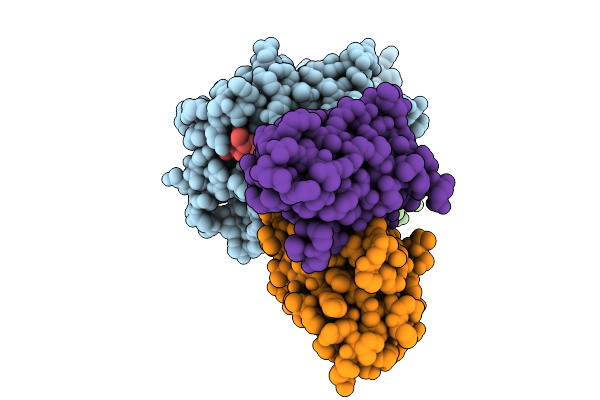
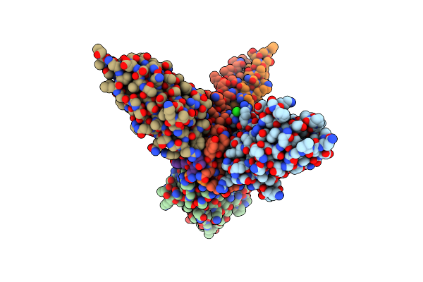
Structure Of The Cross-Hla Supertype Antibody R302 Bound To A Class I Mhc Presenting A Divarasib-Modified Kras-G12C Peptide On Hla-A*02
Organism: Homo sapiens
Method: ELECTRON MICROSCOPY Release Date: 2025-10-08 Classification: PROTEIN BINDING Ligands: A1AWR |
 |
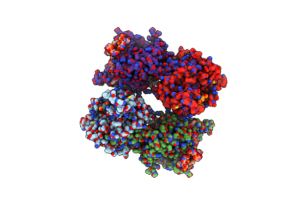
Structure Of The Cross-Hla Supertype Antibody R302 Bound To A Class I Mhc Presenting A Divarasib-Modified Kras-G12C Peptide On Hla-A*03
Organism: Homo sapiens
Method: ELECTRON MICROSCOPY Release Date: 2025-10-08 Classification: PROTEIN BINDING Ligands: A1AWR |
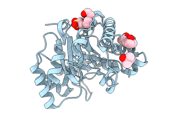 |
Crystal Structure Of S-Nitrosylated Triose Phosphate Isomerase From Chlamydomonas Reinhardtii
Organism: Chlamydomonas reinhardtii
Method: X-RAY DIFFRACTION Release Date: 2025-10-01 Classification: ISOMERASE Ligands: MPD, EPE |
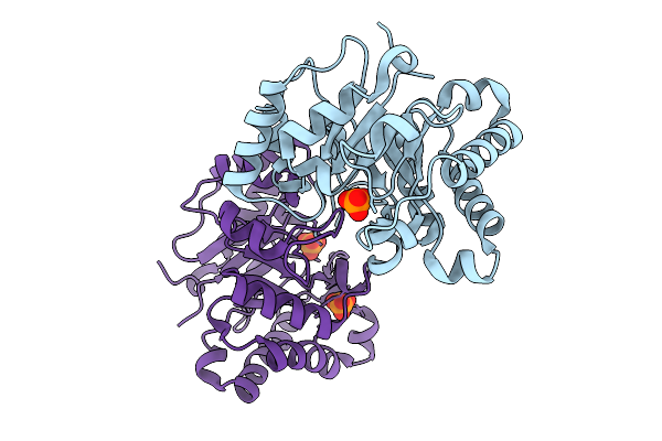 |
Crystal Structure Of C14S Mutant Of Triosephosphate Isomerase From Chamydomonas Reinhardtii
Organism: Chlamydomonas reinhardtii
Method: X-RAY DIFFRACTION Release Date: 2025-10-01 Classification: ISOMERASE Ligands: PO4 |
 |
Organism: Pseudomonas virus pa223
Method: ELECTRON MICROSCOPY Release Date: 2025-08-06 Classification: VIRUS |
 |
Organism: Pseudomonas virus pa223
Method: ELECTRON MICROSCOPY Release Date: 2025-08-06 Classification: VIRUS |
 |
Organism: Pseudomonas virus pa223
Method: ELECTRON MICROSCOPY Release Date: 2025-08-06 Classification: VIRUS |
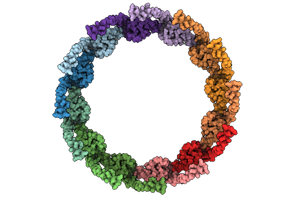 |
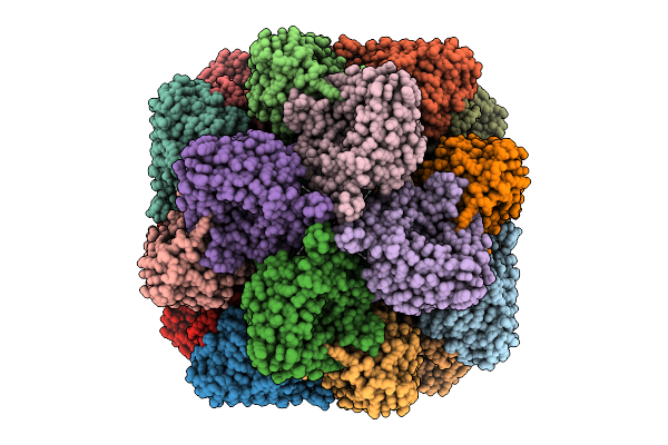
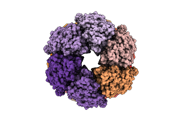
Pseudomonas Phage Pa223 Periplasmic Tunnel (Pre-Ejection, Dodecameric Assembly)
Organism: Pseudomonas virus pa223
Method: ELECTRON MICROSCOPY Release Date: 2025-08-06 Classification: VIRUS |
 |
Organism: Pseudomonas virus pa223
Method: ELECTRON MICROSCOPY Release Date: 2025-08-06 Classification: VIRUS |
 |
Organism: Pseudomonas virus pa223
Method: ELECTRON MICROSCOPY Release Date: 2025-08-06 Classification: VIRUS |
 |
Organism: Pseudomonas virus pa223
Method: ELECTRON MICROSCOPY Release Date: 2025-08-06 Classification: VIRUS |
 |
Hybrid Model Of A Complex Of Brex Proteins Brxb And Pglz From Salmonella Typhimurium
Organism: Salmonella enterica subsp. enterica serovar typhimurium str. d23580
Method: ELECTRON MICROSCOPY Release Date: 2025-07-02 Classification: ANTIMICROBIAL PROTEIN |
 |
Crystal Structure Of Hp1Alpha Chromoshadow Domain In Complex With Kap1 Peptide
Organism: Homo sapiens
Method: X-RAY DIFFRACTION Release Date: 2025-06-11 Classification: TRANSCRIPTION/PEPTIDE Ligands: PGE |
 |
Organism: Homo sapiens
Method: X-RAY DIFFRACTION Release Date: 2025-06-11 Classification: TRANSCRIPTION |
 |
Organism: Lama glama, Severe acute respiratory syndrome coronavirus 2
Method: X-RAY DIFFRACTION Release Date: 2025-05-21 Classification: ANTIVIRAL PROTEIN Ligands: CL |
 |
Organism: Influenza d virus
Method: ELECTRON MICROSCOPY Release Date: 2025-05-14 Classification: VIRAL PROTEIN/RNA |