
Search Count: 41
 |
Organism: Lactiplantibacillus plantarum
Method: X-RAY DIFFRACTION Resolution:1.86 Å Release Date: 2023-04-26 Classification: CELL CYCLE Ligands: SO4, EDO |
 |
Organism: Lactiplantibacillus plantarum
Method: X-RAY DIFFRACTION Resolution:1.95 Å Release Date: 2023-04-26 Classification: CELL CYCLE Ligands: TLA |
 |
Organism: Lactiplantibacillus plantarum
Method: X-RAY DIFFRACTION Resolution:1.94 Å Release Date: 2023-04-26 Classification: CELL CYCLE Ligands: TCE, TLA, GOL |
 |
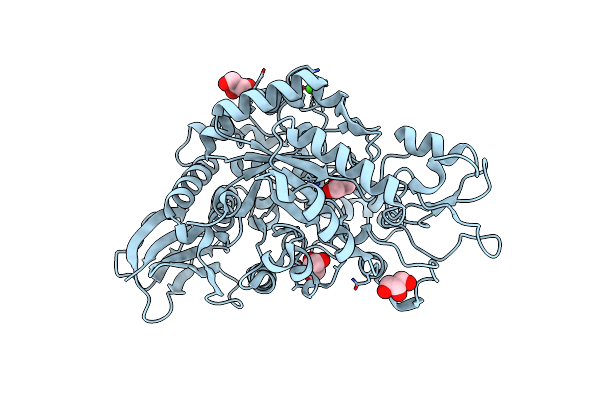
Organism: Lactiplantibacillus plantarum
Method: X-RAY DIFFRACTION Resolution:1.40 Å Release Date: 2023-04-26 Classification: CELL CYCLE Ligands: TLA, GOL, G3P |
 |
Organism: Lactiplantibacillus plantarum subsp. plantarum nc8
Method: X-RAY DIFFRACTION Resolution:2.01 Å Release Date: 2022-08-03 Classification: CYTOSOLIC PROTEIN Ligands: DAL, AMP |
 |
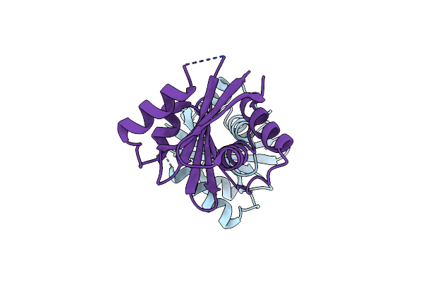
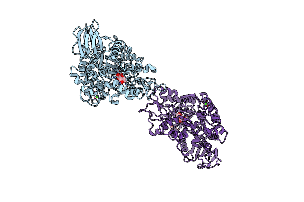
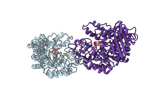
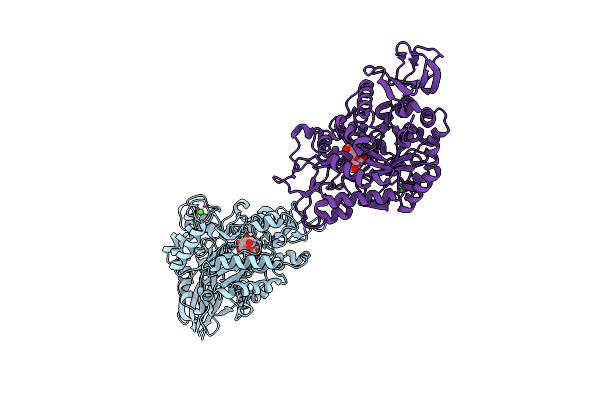
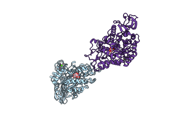
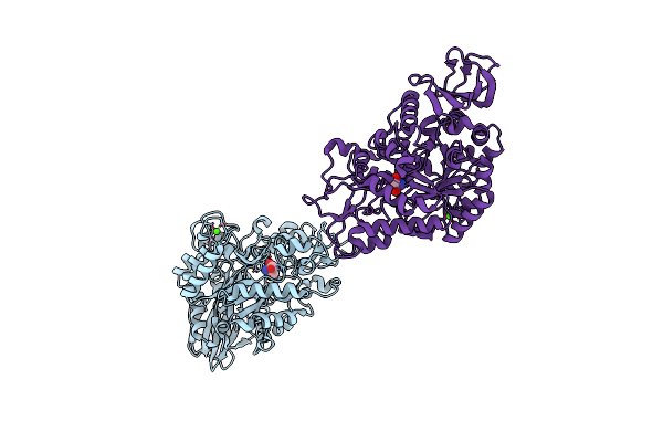
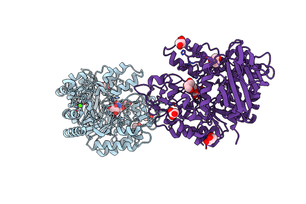
Crystal Structure Of The L. Plantarum Acyl Carrier Protein Synthase (Acps)In Complex With D-Alanyl Carrier Protein (Dltc1)
Organism: Lactiplantibacillus plantarum subsp. plantarum nc8
Method: X-RAY DIFFRACTION Resolution:1.88 Å Release Date: 2022-08-03 Classification: CYTOSOLIC PROTEIN Ligands: PNS, TRS |
 |
Crystal Structure Of The N-Domain Of Hma6, A Copper-Transporting P-Type Atpase
Organism: Arabidopsis thaliana
Method: X-RAY DIFFRACTION Resolution:1.50 Å Release Date: 2017-08-02 Classification: HYDROLASE Ligands: IOD, NH4, CL, EDO |
 |
Crystal Structure Of The N-Domain Of Hma8, A Copper-Transporting P-Type Atpase
Organism: Arabidopsis thaliana
Method: X-RAY DIFFRACTION Resolution:1.75 Å Release Date: 2017-08-02 Classification: HYDROLASE |
 |
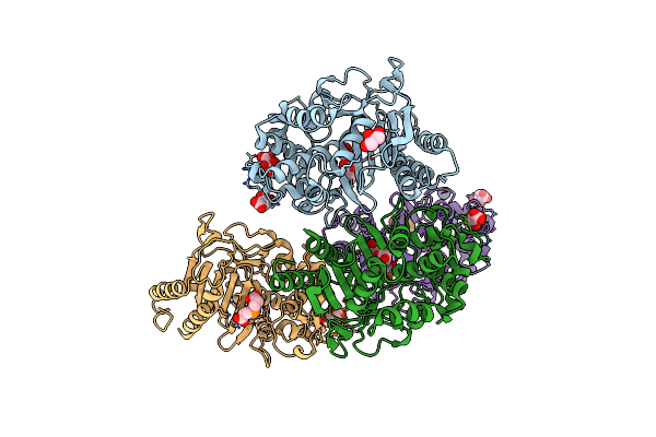
Organism: Rhizobium
Method: X-RAY DIFFRACTION Resolution:1.80 Å Release Date: 2013-09-25 Classification: ISOMERASE Ligands: CA, GLC, GOL |
 |
Organism: Rhizobium
Method: X-RAY DIFFRACTION Resolution:2.00 Å Release Date: 2013-09-25 Classification: ISOMERASE Ligands: GOL, CA, SO4 |
 |
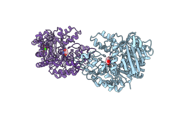
Mutb Inactive Double Mutant D200A-D415N Soaked With Sucrose And Having As Bound Ligands Sucrose In Molecule A And The Reaction Product Trehalulose In Molecule B
Organism: Rhizobium
Method: X-RAY DIFFRACTION Resolution:2.00 Å Release Date: 2013-09-25 Classification: ISOMERASE Ligands: GOL, CA, TEU |
 |
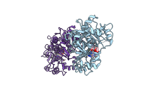
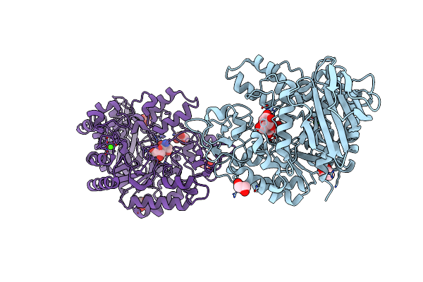
Crystal Structure Of The Trehalulose Synthase Mutb In Complex With Trehalulose
Organism: Rhizobium
Method: X-RAY DIFFRACTION Resolution:1.95 Å Release Date: 2013-09-25 Classification: ISOMERASE Ligands: CA, TEU |
 |
Organism: Rhizobium
Method: X-RAY DIFFRACTION Resolution:2.20 Å Release Date: 2013-09-25 Classification: ISOMERASE Ligands: CA, ISL, GLC |
 |
Crystal Structure Of The Trehalulose Synthase Mutb, Mutant A258V, In Complex With Tris
Organism: Pseudomonas mesoacidophila
Method: X-RAY DIFFRACTION Resolution:2.15 Å Release Date: 2013-08-21 Classification: ISOMERASE Ligands: CA, TRS |
 |
Crystal Structure Of The Trehalulose Synthase Mutant, Mutb D415N, In Complex With Tris
Organism: Pseudomonas mesoacidophila
Method: X-RAY DIFFRACTION Resolution:2.20 Å Release Date: 2013-08-21 Classification: ISOMERASE Ligands: CA, TRS |
 |
Organism: Rhizobium
Method: X-RAY DIFFRACTION Resolution:2.15 Å Release Date: 2013-02-13 Classification: ISOMERASE Ligands: CA, GLC, GOL, TRS |
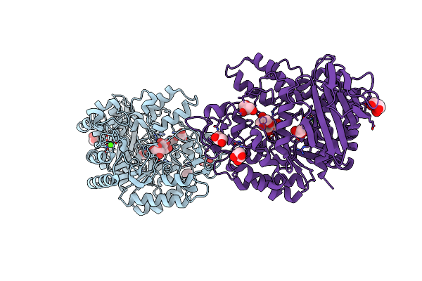 |
Crystal Structure Of The Mutb F164L Mutant From Crystals Soaked With The Substrate Sucrose
Organism: Rhizobium
Method: X-RAY DIFFRACTION Resolution:1.95 Å Release Date: 2013-02-13 Classification: ISOMERASE Ligands: CA, GOL, TRS |
 |
Crystal Structure Of The Mutb F164L Mutant From Crystals Soaked With Trehalulose
Organism: Rhizobium
Method: X-RAY DIFFRACTION Resolution:2.15 Å Release Date: 2013-02-13 Classification: ISOMERASE Ligands: CA, TRS, GOL |
 |
Crystal Structure Of The Mutb F164L Mutant From Crystals Soaked With Isomaltulose
Organism: Rhizobium
Method: X-RAY DIFFRACTION Resolution:2.01 Å Release Date: 2013-02-13 Classification: ISOMERASE Ligands: CA, TRS, GOL |
 |
Crystal Structure Of The Mutb R284C Mutant From Crystals Soaked With The Inhibitor Deoxynojirimycin
Organism: Rhizobium
Method: X-RAY DIFFRACTION Resolution:1.90 Å Release Date: 2013-02-13 Classification: ISOMERASE Ligands: CA, GOL |