
Search Count: 11
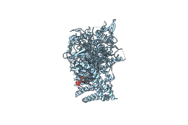 |
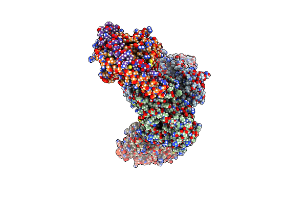
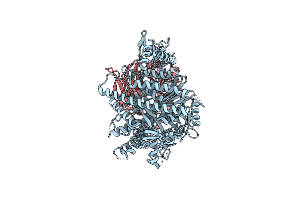
Full-Length P-Rex1 In Complex With Inositol 1,3,4,5-Tetrakisphosphate (Ip4)
Organism: Homo sapiens
Method: ELECTRON MICROSCOPY Release Date: 2024-04-10 Classification: SIGNALING PROTEIN Ligands: 4IP |
 |
Organism: Sus scrofa
Method: ELECTRON MICROSCOPY Release Date: 2024-04-03 Classification: SIGNALING PROTEIN |
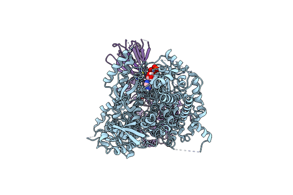 |
Organism: Sus scrofa
Method: ELECTRON MICROSCOPY Release Date: 2024-04-03 Classification: SIGNALING PROTEIN Ligands: ATP |
 |
Organism: Sus scrofa
Method: ELECTRON MICROSCOPY Release Date: 2024-04-03 Classification: SIGNALING PROTEIN Ligands: ADP |
 |
Organism: Sus scrofa, Bos taurus
Method: ELECTRON MICROSCOPY Release Date: 2024-04-03 Classification: SIGNALING PROTEIN Ligands: ADP |
 |
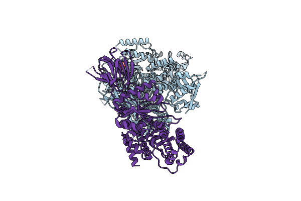
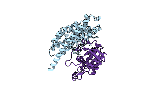
Phosphoinositide Phosphate 3 Kinase Gamma Bound With Adp And Two Gbetagamma Subunits In State 1
Organism: Bos taurus, Sus scrofa
Method: ELECTRON MICROSCOPY Release Date: 2024-04-03 Classification: SIGNALING PROTEIN Ligands: ADP |
 |
Phosphoinositide Phosphate 3 Kinase Gamma Bound With Adp And Two Gbetagamma Subunits In State 2
Organism: Bos taurus, Sus scrofa
Method: ELECTRON MICROSCOPY Release Date: 2024-04-03 Classification: SIGNALING PROTEIN Ligands: ADP |
 |
Organism: Homo sapiens
Method: ELECTRON MICROSCOPY Release Date: 2022-07-06 Classification: SIGNALING PROTEIN |
 |
Organism: Homo sapiens
Method: X-RAY DIFFRACTION Resolution:3.12 Å Release Date: 2020-07-22 Classification: SIGNALING PROTEIN |
 |
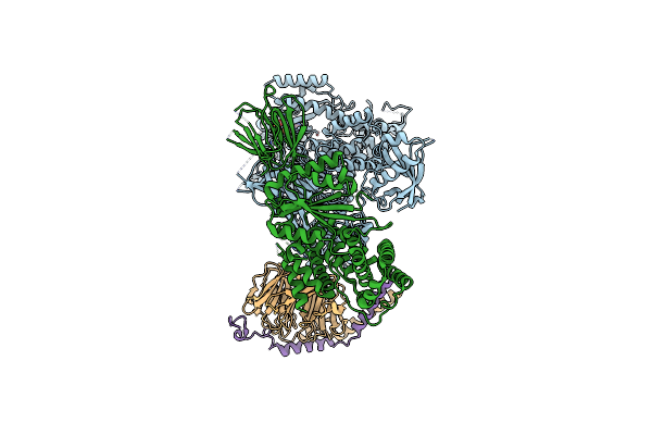
Single Particle Reconstruction Of Phosphatidylinositol (3,4,5) Trisphosphate-Dependent Rac Exchanger 1 Bound To G Protein Beta Gamma Subunits
Organism: Homo sapiens, Bos taurus
Method: ELECTRON MICROSCOPY Release Date: 2019-10-23 Classification: SIGNALING PROTEIN |
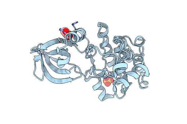 |
Organism: Mycobacterium tuberculosis
Method: X-RAY DIFFRACTION Resolution:2.03 Å Release Date: 2015-02-04 Classification: TRANSFERASE Ligands: GOL, SO4 |