
Search Count: 213
 |
Organism: Homo sapiens
Method: ELECTRON MICROSCOPY Release Date: 2025-10-08 Classification: STRUCTURAL PROTEIN Ligands: ADP, MG, PO4 |
 |
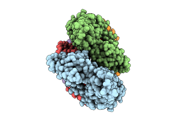
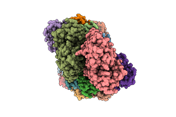
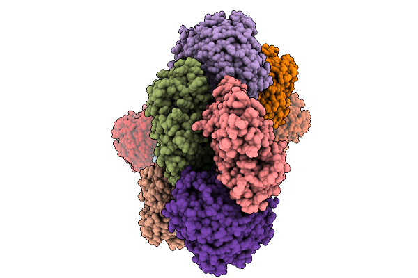
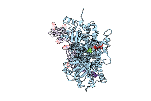
Cryo-Em Structure Of The Actin Filament Bound By A Single Coronin-1B Molecule.
Organism: Homo sapiens
Method: ELECTRON MICROSCOPY Release Date: 2025-10-08 Classification: STRUCTURAL PROTEIN Ligands: ADP, MG, PO4 |
 |
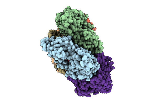
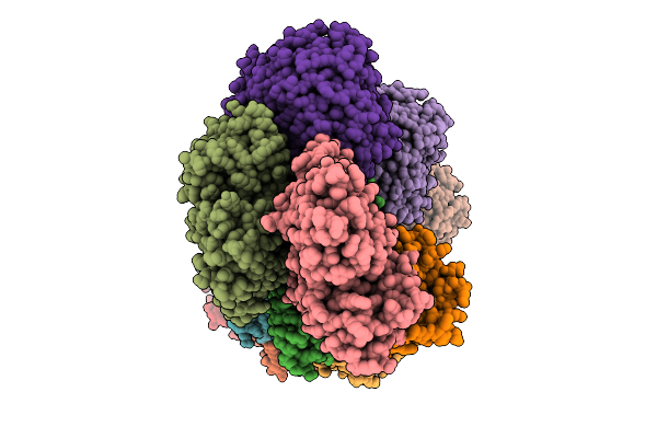
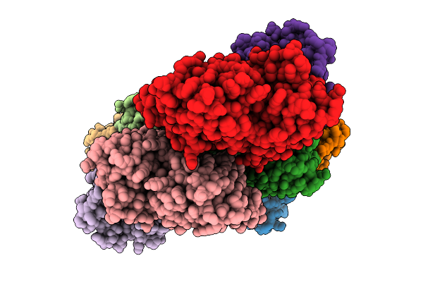
Cryo-Em Structure Of The Fully Coronin-1B-Decorated Actin Filament In The Adp State.
Organism: Homo sapiens
Method: ELECTRON MICROSCOPY Release Date: 2025-10-08 Classification: STRUCTURAL PROTEIN Ligands: ADP, MG |
 |
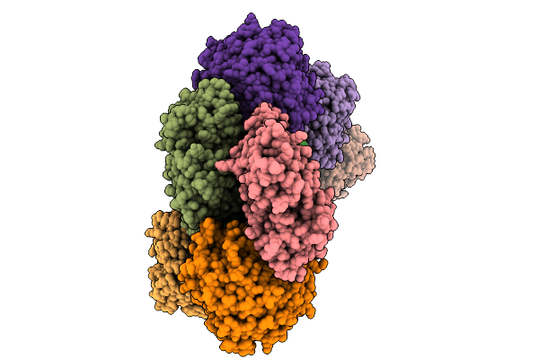
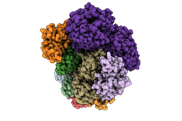
Cryo-Em Structure Of The Fully Coronin-1B-Decorated Actin Filament In The Adp State.
Organism: Homo sapiens
Method: ELECTRON MICROSCOPY Release Date: 2025-10-08 Classification: STRUCTURAL PROTEIN Ligands: ADP, MG |
 |
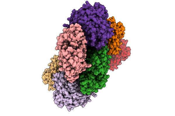
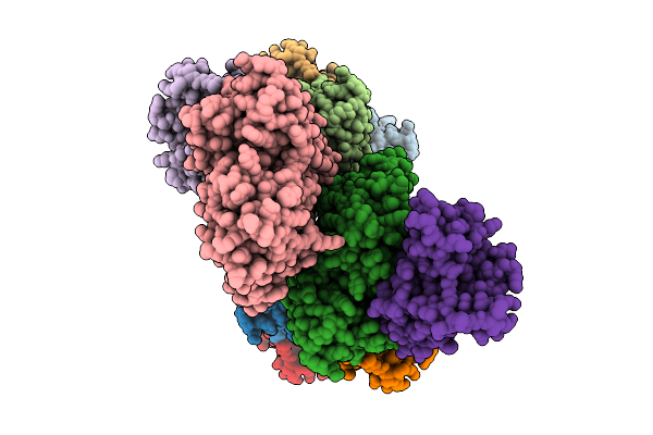
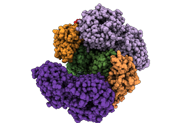
Cryo-Em Structure Of The Fully Cofilin-1-Decorated Actin Filament (Cofilactin)
Organism: Homo sapiens
Method: ELECTRON MICROSCOPY Release Date: 2025-10-08 Classification: STRUCTURAL PROTEIN Ligands: ADP, MG |
 |
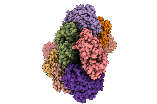
Cryo-Em Structure Of The Actin Filament Hetero-Decorated By Coronin-1 And Cofilin-1 On Separate Actin Strands
Organism: Homo sapiens
Method: ELECTRON MICROSCOPY Release Date: 2025-10-08 Classification: STRUCTURAL PROTEIN Ligands: ADP, MG |
 |
Cryo-Em Structure Of The Actin Filament Hetero-Decorated By Coronin-1 And Cofilin-1, Strand Boundary
Organism: Homo sapiens
Method: ELECTRON MICROSCOPY Release Date: 2025-10-08 Classification: STRUCTURAL PROTEIN Ligands: ADP, MG |
 |
Cryo-Em Structure Of The Cofilactin Filament Core At 2.3 Angstrom Resolution.
Organism: Homo sapiens
Method: ELECTRON MICROSCOPY Release Date: 2025-10-08 Classification: STRUCTURAL PROTEIN Ligands: ADP, MG |
 |
Cryo-Em Structure Of The Coronin-1B-Decorated Actin Filament Bound By One Cofilin-1 Molecule (Crosslinked)
Organism: Homo sapiens
Method: ELECTRON MICROSCOPY Release Date: 2025-10-08 Classification: STRUCTURAL PROTEIN Ligands: ADP, MG |
 |
Organism: Homo sapiens
Method: ELECTRON MICROSCOPY Release Date: 2025-10-08 Classification: STRUCTURAL PROTEIN Ligands: ADP, MG |
 |
Organism: Homo sapiens
Method: ELECTRON MICROSCOPY Release Date: 2025-10-08 Classification: STRUCTURAL PROTEIN Ligands: MG, ADP |
 |
Organism: Homo sapiens
Method: ELECTRON MICROSCOPY Release Date: 2025-10-08 Classification: STRUCTURAL PROTEIN Ligands: MG, ADP |
 |
Organism: Xenopus laevis, Homo sapiens, Synthetic construct
Method: ELECTRON MICROSCOPY Release Date: 2025-08-06 Classification: IMMUNE SYSTEM Ligands: NAG |
 |
Organism: Xenopus laevis, Synthetic construct
Method: ELECTRON MICROSCOPY Release Date: 2025-08-06 Classification: NUCLEAR PROTEIN |
 |
Organism: Xenopus laevis, Synthetic construct, Homo sapiens
Method: ELECTRON MICROSCOPY Release Date: 2025-08-06 Classification: IMMUNE SYSTEM Ligands: NAG, HEM |
 |
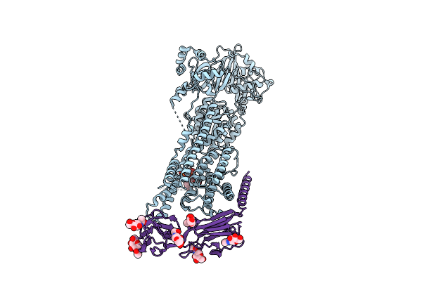
Native Dimeric Myeloperoxidase Bound To Nucleosome Core Particle; Composite Map
Organism: Xenopus laevis, Synthetic construct, Homo sapiens
Method: ELECTRON MICROSCOPY Release Date: 2025-08-06 Classification: IMMUNE SYSTEM Ligands: CL, HEM, NAG, CA |
 |
Native Dimeric Myeloperoxidase Bound To Nucleosome Core Particle, Intermediate State; Composite Map
Organism: Xenopus laevis, Synthetic construct, Homo sapiens
Method: ELECTRON MICROSCOPY Release Date: 2025-08-06 Classification: IMMUNE SYSTEM Ligands: CL, HEM, NAG, CA |
 |
Organism: Mus musculus
Method: ELECTRON MICROSCOPY Release Date: 2025-08-06 Classification: MEMBRANE PROTEIN Ligands: MG, KXP, BEF, NAG |
 |
Cryo-Em Structure Of Mouse Pmca-Nptn Complex Captured In E1 State Without Calcium
Organism: Mus musculus
Method: ELECTRON MICROSCOPY Release Date: 2025-08-06 Classification: MEMBRANE PROTEIN Ligands: KXP, NAG |
 |
Organism: Mus musculus
Method: ELECTRON MICROSCOPY Release Date: 2025-08-06 Classification: MEMBRANE PROTEIN Ligands: ANP, MG, KXP, NAG |