
Search Count: 25
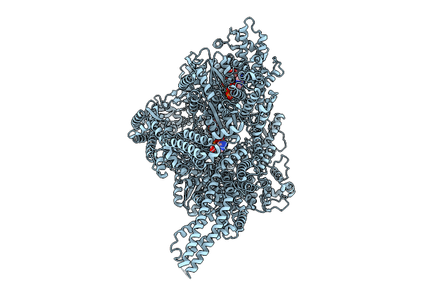 |
Organism: Sus scrofa
Method: ELECTRON MICROSCOPY Release Date: 2025-09-10 Classification: MOTOR PROTEIN Ligands: ADP, ATP, ANP, MG |
 |
Organism: Sus scrofa
Method: ELECTRON MICROSCOPY Release Date: 2025-09-10 Classification: MOTOR PROTEIN Ligands: ADP, ATP, MG, ANP |
 |
Organism: Sus scrofa
Method: ELECTRON MICROSCOPY Release Date: 2025-09-10 Classification: MOTOR PROTEIN Ligands: ZN, MG |
 |
Organism: Sus scrofa
Method: ELECTRON MICROSCOPY Release Date: 2025-09-10 Classification: MOTOR PROTEIN Ligands: ADP, ATP, ZN |
 |
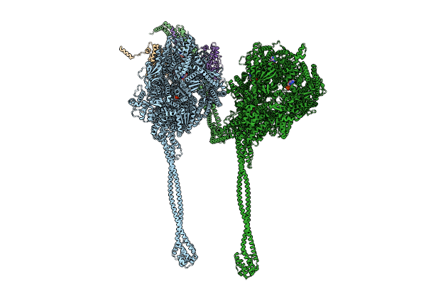
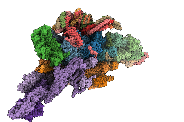
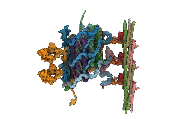
Structure Of Dynactin, Dynein Tail With One Bicdr From Dynein-Dynactin-Bicdr On Microtubules
Organism: Mus musculus, Sus scrofa
Method: ELECTRON MICROSCOPY Release Date: 2025-09-10 Classification: MOTOR PROTEIN Ligands: ADP, ATP, ZN |
 |
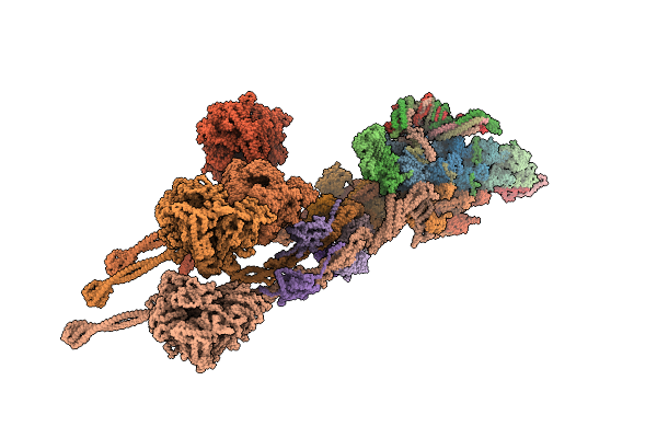
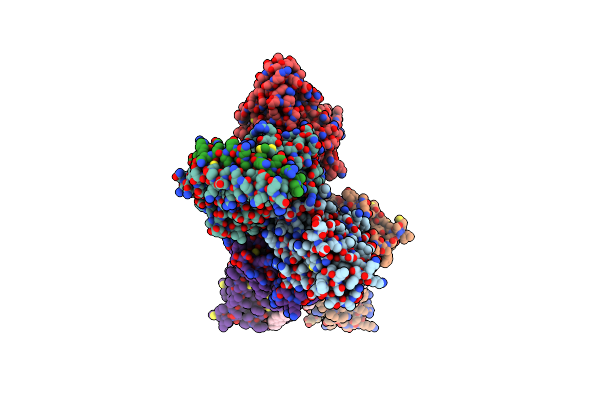
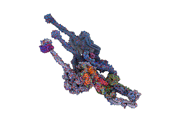
Structure Of Dynactin, Dynein Tail With Two Bicdr From Dynein-Dynactin-Bicdr On Microtubules
Organism: Mus musculus, Sus scrofa
Method: ELECTRON MICROSCOPY Release Date: 2025-09-10 Classification: MOTOR PROTEIN Ligands: ADP, ATP, ZN |
 |
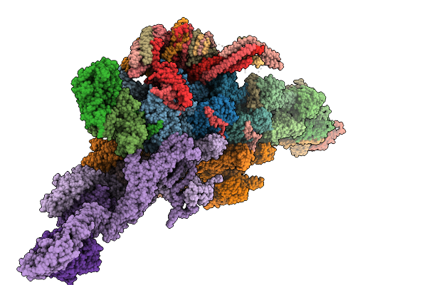
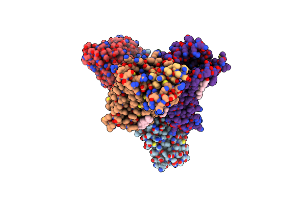
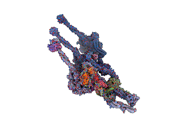
Structure Of Dynactin, Dynein Tail With Trak2 From Dynein-Dynactin-Trak2 On Microtubules
Organism: Mus musculus, Sus scrofa
Method: ELECTRON MICROSCOPY Release Date: 2025-09-10 Classification: MOTOR PROTEIN Ligands: ADP, ATP, ZN |
 |
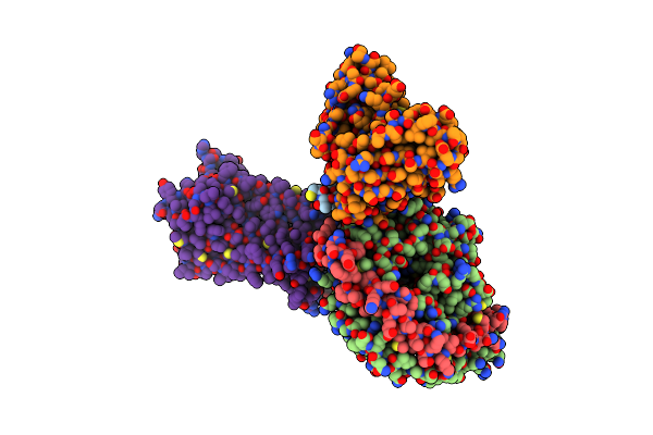
Organism: Homo sapiens, Rattus norvegicus, Bos taurus, Synthetic construct
Method: ELECTRON MICROSCOPY Release Date: 2022-11-09 Classification: SIGNALING PROTEIN Ligands: 7V7, CLR |
 |
Organism: Homo sapiens, Rattus norvegicus, Bos taurus, Synthetic construct
Method: ELECTRON MICROSCOPY Release Date: 2022-11-09 Classification: SIGNALING PROTEIN Ligands: MOI, CLR |
 |
Organism: Homo sapiens, Rattus norvegicus, Bos taurus, Synthetic construct
Method: ELECTRON MICROSCOPY Release Date: 2022-11-09 Classification: SIGNALING PROTEIN Ligands: WH2 |
 |
Organism: Homo sapiens, Rattus norvegicus
Method: ELECTRON MICROSCOPY Release Date: 2022-11-09 Classification: SIGNALING PROTEIN Ligands: WH9, CLR |
 |
Organism: Homo sapiens, Rattus norvegicus, Bos taurus, Synthetic construct
Method: ELECTRON MICROSCOPY Release Date: 2022-11-09 Classification: SIGNALING PROTEIN Ligands: 8QY, CLR |
 |
Organism: Homo sapiens, Rattus norvegicus, Bos taurus, Synthetic construct
Method: ELECTRON MICROSCOPY Release Date: 2022-11-09 Classification: SIGNALING PROTEIN Ligands: ETA |
 |
Organism: Chlamydomonas reinhardtii
Method: ELECTRON MICROSCOPY Release Date: 2022-05-18 Classification: STRUCTURAL PROTEIN Ligands: ADP, GDP, GTP |
 |
Organism: Chlamydomonas reinhardtii
Method: ELECTRON MICROSCOPY Release Date: 2022-05-18 Classification: STRUCTURAL PROTEIN Ligands: GDP, GTP |
 |
Structure Of Outer-Arm Dyneins Bound To Microtubule With Microtubule Binding State 1(Mtbs-1)
Organism: Tetrahymena thermophila
Method: ELECTRON MICROSCOPY Resolution:4.00 Å Release Date: 2021-09-29 Classification: STRUCTURAL PROTEIN Ligands: ADP, ATP, MG |
 |
Structure Of Outer-Arm Dynein Bound To Microtubule Doublet In Microtubule Binding State 2 (Mtbs-2)
Organism: Tetrahymena thermophila
Method: ELECTRON MICROSCOPY Release Date: 2021-09-29 Classification: MOTOR PROTEIN Ligands: ADP, ATP, MG |
 |
Organism: Tetrahymena thermophila
Method: ELECTRON MICROSCOPY Release Date: 2021-09-29 Classification: MOTOR PROTEIN Ligands: ADP, ATP, MG |
 |
Organism: Tetrahymena thermophila
Method: ELECTRON MICROSCOPY Release Date: 2021-09-29 Classification: STRUCTURAL PROTEIN Ligands: GTP, MG, GDP |
 |
Organism: Tetrahymena thermophila
Method: ELECTRON MICROSCOPY Resolution:4.50 Å Release Date: 2021-09-29 Classification: STRUCTURAL PROTEIN Ligands: GTP, MG, GDP |